
Scalp: Difference between revisions
→Innervation: +mnemonic, heading |
Removing incorrect ref for nerve supply |
||
| Line 46: | Line 46: | ||
==Innervation== |
==Innervation== |
||
Innervation is the connection of nerves to the scalp; the sensory and motor nerves innervating the scalp. The scalp is innervated by the following:<ref> |
Innervation is the connection of nerves to the scalp; the sensory and motor nerves innervating the scalp. The scalp is innervated by the following:<ref name="LHC"></ref> |
||
* [[Supratrochlear nerve]] and the [[supraorbital nerve]] from the [[ophthalmic division]] of the [[trigeminal nerve]] |
* [[Supratrochlear nerve]] and the [[supraorbital nerve]] from the [[ophthalmic division]] of the [[trigeminal nerve]] |
||
* [[Greater occipital nerve]] (C2) posteriorly up to the vertex |
* [[Greater occipital nerve]] (C2) posteriorly up to the vertex |
||
Revision as of 15:47, 15 December 2009

The scalp is the anatomical area bordered by the face anteriorly and the neck to the sides and posteriorly.
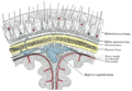
Layers
It is usually described as having five layers, which can be remembered with the mnemonic "SCALP":[1].
- S: The skin on the head from which head hair grows. It is richly supplied with blood vessels.
- C: Connective tissue. A thin layer of fat and fibrous tissue lies beneath the skin.
- A: The aponeurosis called epicranial aponeurosis (or galea aponeurotica) is the next layer. It is a tough layer of dense fibrous tissue which runs from the frontalis muscle anteriorly to the occipitalis posteriorly.
- L: The loose areolar connective tissue layer provides an easy plane of separation between the upper three layers and the pericranium. In scalping the scalp is torn off through this layer. It also provides a plane of access in craniofacial surgery and neurosurgery. This layer is sometimes referred to as the "Danger Zone" because of the ease by which infectious agents can spread through it to emissary veins which then drain into the cranium. The loose areolar tissue in this layer is made up of random collagen I bundles, collagen III. It contains the majors blood vessels of the scalp, which bleed profusely upon injury, partly due to the absence of venous valves found in the circulation below the neck. It will also be rich in glycosaminoglycans (GAGs) and will be constituted of more matrix than fibers.
- P: The pericranium is the periosteum of the skull bones and provides nutrition to the bone and the capacity for repair. It may be lifted from the bone to allow removal of bone windows (craniotomy).
The clinically important layer is the aponeurosis. Scalp lacerations through this layer mean that the "anchoring" of the superficial layers is lost and gaping of the wound occurs; this requires suturing. This can be achieved with simple or vertical matress sutures using a non-absorbable material, which are subsequently removed at around days 7-10.
Blood supply
The blood supply of the scalp is via five pairs of arteries, three from the external carotid and two from the internal carotid:
- internal carotid
- the supratrochlear artery to the midline forehead. supratrochlear artery is a branch of ophthalmic branch of the internal carotid artery.
- the supraorbital artery to the lateral forehead and scalp as far up as the vertex. supraorbital artery is a branch of ophthalmic branch of the internal carotid artery.
- external carotid
- the superficial temporal artery which gives frontal and parietal branches to supply much of the scalp
- the occipital artery which runs from posteriorly to supply much of the back of the scalp.
- the posterior auricular artery , a branch of the external carotid artery , ascends behind the auricle to supply the scalp above and behind the auricle.
Innervation
Innervation is the connection of nerves to the scalp; the sensory and motor nerves innervating the scalp. The scalp is innervated by the following:[2]
- Supratrochlear nerve and the supraorbital nerve from the ophthalmic division of the trigeminal nerve
- Greater occipital nerve (C2) posteriorly up to the vertex
- Lesser occipital nerve (C3) behind the ear.
- Zygomaticotemporal nerve from the maxillary division of the trigeminal nerve supplying the hairless temple
- Auriculotemporal nerve from the mandibular division of the trigeminal nerve
The innervation of scalp can be remembered using the mnemonic, "Z-GLASS" for, Zygomaticotemporal nerve, Greater occipital nerve, Lesser occipital nerve, Auriculotemporal nerve, Supratrochlear nerve and Supraorbital nerve.[2]
Lymphatic drainage
There are no lymph nodes in the scalp; lymphatic drainage is to the pre- and post-auricular nodes.
Role in aesthetics
The scalp plays an important role in the aesthetics of the face. Androgenic alopecia, or male pattern hair loss, is a common cause of concern to men. It may be treated by medication (eg finasteride, minoxidil) or hair transplantation with variable success. If the scalp is heavy and loose, a common change with aging, the forehead may be low, heavy and deeply lined. The brow lift procedure aims to address these concerns.
Pathology
The scalp is a common site for the development of tumours including:
- epidermoid cyst
- pilar cyst
- actinic keratosis and squamous cell carcinoma
- basal cell carcinoma
- merkel cell tumours
Scalp conditions
- Dandruff -- A common problem due to the excessive shedding of dead skin cells from the scalp
- Seborrhoeic dermatitis -- a skin disorder causing scaly, flaky, itchy, red skin
- Cutis verticis gyrata -- A descriptive term for a rare deformity of the scalp.
- Head lice
See also
- Trichology -- the scientific study of hair and scalp
- Trichodynia -- burning scalp syndrome
- Scalping -- the act of removing the scalp, usually with the hair, as a portable proof or trophy of prowess in war.
Additional images
-
 Diagrammatic section of scalp.

References
External links
- Histology image: 08601ooa – Histology Learning System at Boston University - "Integument: scalp, transverse"
- Histology image: 08801ooa – Histology Learning System at Boston University - "Integument: scalp"
- lesson1 at The Anatomy Lesson by Wesley Norman (Georgetown University)
- http://www.dartmouth.edu/~humananatomy/figures/chapter_47/47-1.HTM