
Fibroadenoma
| Fibroadenoma | |
|---|---|
| Specialty | Oncology, gynaecology |
Fibroadenomas of the breast, are lumps composed of fibrous and glandular tissue. Because breast cancer can also appear as a lump, doctors may recommend a tissue sample (biopsy) to rule out cancer in older patients. Unlike typical lumps from breast cancer, fibroadenomas are easy to move, with clearly defined edges.[1][2]
Fibroadenomas are sometimes called breast mice or a breast mouse owing to their high mobility in the breast.[3]
Signs and symptoms
The typical case is the presence of a painless, firm, solitary, mobile, slowly growing lump in the breast of a woman of childbearing years.[2][4][5]
In the male breast, fibroepithelial tumors are very rare, and are mostly phyllodes tumors. Exceptionally rare case reports exist of fibroadenomas in the male breast, however these cases may be associated with antiandrogen treatment.[6]
Diagnosis

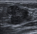
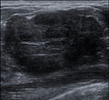
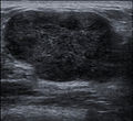
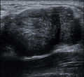
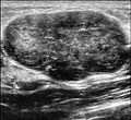
A fibroadenoma is usually diagnosed through clinical examination, ultrasound or mammography, and often a needle biopsy sample of the lump.[4]
Etiology and epidemiology
Fibroadenomas arise in the terminal duct lobular unit of the breast.[citation needed] They are the most common breast tumor in adolescent women. They also occur in a small number of post-menopausal women. Their incidence declines with increasing age, and, in general, they appear before the age of thirty years. Fibroadenomas are partially hormone-dependent and frequently regress after menopause. They are hypovascular compared to typical (especially malignant) neoplasms.[2][7][5]
Higher intake of fruits and vegetables, higher number of live births, use of oral contraceptives and moderate exercise are associated with lower frequency of fibroadenomas. [8]
Pathology

Cytology
The diagnostic findings on needle biopsy consist of abundant stromal cells, which appear as bare bipolar nuclei, throughout the aspirate; sheets of fairly uniform-size epithelial cells that are typically arranged in either an antler-like pattern or a honeycomb pattern. These epithelial sheets tend to show typical metachromatic blue staining on DiffQuick staining. Foam cells and apocrine cells may also be seen, although these are less diagnostic features.[4][7] The gallery images below demonstrate these features.
Macroscopic
Approximately ninety percent of fibroadenomas are less than three centimetres in diameter. The vast majority of the remaining ten percent that are four centimetres or larger occur mostly in women under twenty years of age. The tumor is round or ovoid, elastic, and nodular, and has a smooth surface. The cut surface usually appears homogenous and firm, and is grey-white or tan in colour. The pericanalicular type (hard) has a whorly appearance with a complete capsule, while the intracanalicular type (soft) has an incomplete capsule.[7]
Microscopic
Fibroadenoma of the breast is a benign tumor composed of two elements : epithelium and stroma. It is nodular and encapsulated, included in breast. The epithelial proliferation appears in a single terminal ductal unit and describes duct-like spaces surrounded by a fibroblastic stroma. Depending on the proportion and the relationship between these two components, there are two main histological features : intracanalicular and pericanalicular. Often, both types are found in the same tumor. A) Intracanalicular fibroadenoma : stromal proliferation predominates and compresses the ducts, which are irregular, reduced to slits. B) Pericanalicular fibroadenoma : fibrous stroma proliferates around the ductal spaces, so that they remain round or oval, on cross section. The basement membrane is intact [9] The gallery image below demonstrates both morphological subtypes.
Treatment
Most fibroadenomas are left in situ and monitored by a doctor, or the patient in question. Some are treated by surgical excision. They are removed with a small margin of normal breast tissue if the preoperative clinical investigations are suggestive of the diagnosis. A small amount of normal tissue must be removed in case the lesion turns out to be a phyllodes tumour on microscopic examination.[7][10]
Because needle biopsy is often a reliable diagnostic investigation, some doctors may decide not to operate to remove the lesion, and instead opt for clinical follow-up to serially observe the lesion over time using clinical examination and mammography to determine the rate of growth, if any, of the lesion. A growth rate of less than sixteen percent per month in women under fifty years of age, and a growth rate of less than thirteen percent per month in women over fifty years of age have been published as safe growth rates for continued non-operative treatment and clinical observation.[11]
Some fibroadenomas respond to treatment with ormeloxifene.[12]
Fibroadenomas have not been shown to recur following complete excision or transform into phyllodes tumours following partial or incomplete excision.[7]
There are also natural treatments being touted to diminish fibroadenomas, such as Fibrosolve, but no definite studies have been made as to prove their effectiveness.
Ultrasound Treatment
Europe recently recognized an alternative treatment that is called echotherapy and that uses high-intensity focused ultrasound to treat breast fibroadenoma.[13]. This method is non-invasive and relies on tissue heating to detroy fibroadenoma cells. Focused ultrasounds have been used for a long time in the treatment of differents tumors such as prostate, liver or uterus, where they proved their efficacy.[14][15][16][17].
A French company, Theraclion, develops an echotherapy device for benign tumors treatment especially breast fibroadenoma.
Cryoablation Treatment
The FDA has approved cryoablation (the use of extreme cold to destroy tissue) of a fibroadenoma as a safe, effective and minimally-invasive alternative to open surgical removal.[18] In the procedure, ultrasound imaging is used to guide a probe into the mass of breast tissue. Extremely cold temperatures are then used to destroy the abnormal cells,[19] and over time the cells are reabsorbed into the body. The procedure can be performed in an office setting with local anesthesia only, and leaves substantially less scarring than open surgical procedures.[19]
The American Society of Breast Surgeons recommends the following criteria to establish a patient as a candidate for cryoablation of a fibroadenoma:[18]
- The lesion must be sonographically visible.
- The diagnosis of fibroadenoma must be confirmed histologically.
- Lesions should be less than 4 cm in diameter.
Fibroadenoma images
-
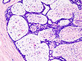
 Fibroadenoma Histology (H&E). The image demonstrates intracanalicular morphology (top right) and pericanalicular morphology (bottom left)
 -
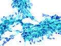
 Fibroadenoma, Fine Needle Aspiration Biopsy (Giemsa or DiffQuickTM stain). The image shows abundant bare bipolar stromal nuclei surrounding sheets of metachromatic epithelial cells.
 -
 Fibroadenoma, Fine Needle Aspiration Biopsy (Papanicolou stain). The image shows a sheet of epithelial cells in the typical antler pattern.
 -
 Histopathologic image of breast fibroadenoma. Core needle biopsy. Hematoxylin & eosin stain.
 -

-

-

-

-

-
-

-

-
-

-
-

-

-
-

-
-
.jpg)
_DG_stain.jpg)
_PAP_stain.jpg)
.jpg)


Video on Fibroadenoma
- Fibroadenoma Video
- The Doctors Show: Fibroadenoma Cryoabltaion on YouTube
External Links
- Theraclion EchoPulse Ultrasound Therapy for Breast Fibroadenomas Cleared in EU
- Theraclion official website
References
- ^ Fibroadenomas at Merck Manual of Diagnosis and Therapy Home Edition
- ^ a b c Tavassoli, F.A., Devilee, P., ed. (2003). World Health Organization Classification of Tumours: Pathology & Genetics: Tumours of the breast and female genital organs. Lyon: IARC Press. ISBN 92-832-2412-4.
{{cite book}}: CS1 maint: multiple names: editors list (link) - ^ Scott-Conner, edited by Carol E.H. (2010). Breast surgery office management and surgical techniques. New York: Springer. p. 71. ISBN 978-1-4419-6075-7.
{{cite book}}:|first=has generic name (help); Unknown parameter|coauthors=ignored (|author=suggested) (help) - ^ a b c DeMay, M. Practical Principles of Cytopathology (Revised ed.). ASCP Press. p. 2007. ISBN 0-89189-549-3.
- ^ a b Pathology Outlines Website. [1] Accessed 12 February 2009.
- ^ Shin SJ, Rosen PP (2007). "Bilateral presentation of fibroadenoma with digital fibroma-like inclusions in the male breast". Arch. Pathol. Lab. Med. 131 (7): 1126–9. doi:10.1043/1543-2165(2007)131[1126:BPOFWD]2.0.CO;2. PMID 17617003.
{{cite journal}}: Unknown parameter|month=ignored (help) - ^ a b c d e Rosen, PP. Rosen's Breast Pathology (3rd ed.). ISBN 978-0-7817-7137-5.
- ^ Attention: This template ({{cite pmid}}) is deprecated. To cite the publication identified by PMID 20484549, please use {{cite journal}} with
|pmid=20484549instead. - ^ "Fibroadenoma of the breast". Retrieved 15 December 2007.
- ^ Rosai, J. (2004). Rosai and Ackerman's Surgical Pathology (9th ed.). ISBN 0-323-01342-2.
- ^
Gordon PB, Gagnon FA, Lanzkowsky L (2003). "Solid breast masses diagnosed as fibroadenoma at fine-needle aspiration biopsy: acceptable rates of growth at long-term follow-up". Radiology. 229 (1): 233–8. doi:10.1148/radiol.2291010282. PMID 14519878.
{{cite journal}}: Unknown parameter|month=ignored (help)CS1 maint: multiple names: authors list (link) - ^ Attention: This template ({{cite pmid}}) is deprecated. To cite the publication identified by PMID 17431715, please use {{cite journal}} with
|pmid=17431715instead. - ^ http://www.medgadget.com/2012/12/theraclion-echopulse-ultrasound-therapy-for-breast-fibroadenomas-cleared-in-eu.html
- ^ Jolesz FA, Hynynen K, McDannold N, Tempany C. MR imaging-controlled focused ultrasound ablation: a noninvasive image-guided surgery. Magn Reson Imaging Clin N Am 2005;13(3):545-60.
- ^ Gombos EC, Kacher DF, Furusawa H, Namba K. Breast focused ultrasound surgery with magnetic resonance guidance. Top Magn Reson Imaging 2006;17(3):181-8.
- ^ Hynynen K, Pomeroy O, Smith DN, Huber PE, McDannold NJ, Kettenbach J, Baum J, Singer S, Jolesz FA. MR imaging-guided focused ultrasound surgery of fibroadenomas in the breast: a feasibility study. Radiology 2001;219:176-185.
- ^ Marret H, Bleuzen A, Guérin A, Lauvin-Gaillard MA, Herbreteau D, Patat F, Tranquart F. French first results using magnetic resonance-guided focused ultrasound for myoma treatment. Gynecol Obstet Fertil. 2011 Jan;39(1):12-20. Epub 2010 Dec 24.
- ^ a b Management of Fibroadenomas of the Breast
- ^ a b WebMD — Cryotherapy Shrinks Benign Breast Lumps
.