Chromatophore
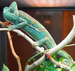
Chromatophore is the collective term for pigment containing and light reflecting cells found in amphibians, fish, reptiles, crustaceans and cephalopods. Subclassed as xanthophores, erythrophores, iridophores, leucophores, melanophores and cyanophores according to their hue, chromatophores are largely responsible for generating skin colour in poikilothermic animals. The translocation of pigment within the cells is the mechanism through which some species, like chameleons and octopus, can rapidly change colour. The mammalian and avian equivalent of the chromatophore cell is the melanocyte.
Classification
Chromforo was first used to describe invertebrate pigment cells in 1819 and the term chromatophore (Greek: khrōma = "colour", phoros = "bearing") later adopted as a name for the pigment bearing cells of cold blooded vertebrates and cephalopods (in contrast to the chromato-cytes found in mammals and birds). By the 1960s, sufficient understanding of the structure and colour of chromatophores was available to sub-classify them according to appearance and, despite subsequent studies revealing the biochemical nature of the pigments within chromatophore types, this classification system persists. [1]
Xanthophores and erythrophores
Originally termed lipophores due to their fat-soluble content, chromatophores that contain large amounts of yellow pteridine pigments were renamed xanthophores and those with an excess of red/orange carotinoids termed erythrophores. [1] Soon after, it was discovered that pterinosome and carotinoid vesicles are found within the same cell, and that the manifest colour depends on the ratio of red and yellow pigments. [2] Thus the distinction between these chromatophore types is essentially arbitrary.

Iridophores and leucophores
Biochromes, such as pteridines and carotinoids, selectively absorb a part of the visual spectrum that makes up white incident light, while they let the other wavelengths pass and reach the eye of the observer. Not all colours are generated in this manner, however. Some, most notably blues and greens, are generated by the scattering, interference and diffusion of light by colourless crystalline structures called schemochromes.
Iridophores are lower vertebrate pigment cells that reflect light using plates of crystalline guanine schemochromes.[3] When illuminated they generate iridescent colours due to the diffraction of light within the stacked plates. Orientation of the schemochrome determines the nature of the structural colour observed.[4] By using biochromes as filters, iridophores mediate an optical effect known as Tyndall or Rayleigh scattering, producing bright blue or green colours that are not modified by the angle of vision.[5]
A related type of chromatophore, the leucophore, is found in some fish species. Like iridophores, they utilize crystalline purines to reflect light, providing the bright white colour seen in some fish. As with xanthophores and erythrophores, the distinction between iridophores and leucophores in fish is not always obvious, but generally iridophores are considered to generate iridescent or metallic colours while leucophores produce structural white hues.[5]
Melanophores
The most widely studied chromatophore, due both to its extensive taxonomic distribution and apparent colour, is the melanophore. Eumelanin, the biochrome found in melanophores, is generated from tyrosine in a series of catalysed chemical reactions.[6] This type of melanin, when packaged in vesicles and distributed throughout the cell, appears black or dark brown, due to its light absorbing qualities. In some amphibian species, however, there are other pigments packaged alongside eumelanin. For example, a novel deep red coloured pigment called was identified in the melanophores of phyllomedusine frogs.[7] This was subsequently identified as pterorhodin, a pterodine dimer that accumulates around eumelanin. While it is likely that other species have complex melanophore pigments, it is nevertheless true that the majority of melanophores studied to date contain eumelanin exclusively.

Cyanophores
In 1995 it was demonstrated that the vibrant blue colours of mandarin fish are not structural in nature. Instead, a cyan biochrome of unknown chemical nature is responsible.[5] This pigment, found within fibrous vesicles in at least two species callionymid fish, is highly unusual in the animal kingdom, as all other blue colourings thus far investigated are schemochromatic. Therefore a novel chromatophore type, the cyanophore, was proposed. Although cyanophores are unusual in their taxonomic restriction, there may be other unusual chromatophore types in lesser-studied fish and amphibians. Indeed, bright coloured chromatophores with undefined pigments have been observed in both poison dart frogs and glass frogs.[8]
Pigment translocation
Many species have the ability to translocate the pigment inside chromatophores, resulting in an apparent change in colour. This process, known as physiological colour change, is most widely studied in melanophores, as melanin is the darkest and most visible pigment.

In most species with a relatively thin dermis, the dermal melanophores tend to be flat and cover a large surface area. However, in animals with thick dermal layers, such as adult reptiles, dermal melanophores often form three-dimensional units with other chromatophores. These dermal chromatophore units (DCU) consist of an uppermost xanthophore or erythrophore layer, then an iridophore layer, and finally a basket-like melanophore layer with processes covering the iridophores.[9]
Both types of dermal melanophores are extremely important in physiological colour change. Flat dermal melanophores will often overlay other chromatophores so when the pigment is dispersed throughout the cell the skin appears dark. When the pigment is aggregated towards the centre of the cell, the pigments in other chromatophores are exposed to light and thus the skin takes on their hue. Similarly, after melanin aggregation in DCUs, the skin appears green through xanthophore (yellow) filtering of scattered (blue) light from the iridophore layer. On the dispersion of melanin, the light is no longer scattered and the skin appears dark. As the other biochromatic chomatophores are also capable of pigment translocation, by making good use of the divisional effect animals with multiple chromatophore types can generate a spectacular array of skin colours.[10],[11]
The control and mechanics of rapid pigment translocation has been well studied in a multitude of species, particularly amphibians and teleost fish. [12],[5] It has been demonstrated that the process can be under hormonal, neuronal control or both. Neurochemicals that are known to translocate pigment include noradrenaline, via its alpha2-adrenoceptor on the surface on melanophores.[13] The primary hormones involved in regulating translocation appear to be the melanocortins, melatonin and melanin concentrating hormone (MCH), that are produced mainly in the pituitary, pineal gland and hypothalamus respectively, though may also be generated in a paracrine fashion by peripheral tissues. At the surface of the melanophore these peptides have been shown to activate specific G protein coupled receptors that, in turn, transduce the signal into the cell. Melanocortins result in the dispersion of pigment, while melatonin and MCH results in aggregation.[14] Numerous melanocortin, MCH and melatonin receptors have been identified in fish [15] and frogs [16], including the orthologue of MC1R [17], a melanocortin receptor known to regulate skin and hair colour in humans.[18] Inside the cell, cyclic adenosine monophosphate (cAMP) has been shown to be an important second messenger of pigment translocation. Through a mechanism not yet fully understood, cAMP influences other proteins to drive molecular motors carrying melanosomes along both microtubules and microfilaments.[19],[20],[21]
Background adaptation
Most fish, reptiles and amphibians animals undergo physiological colour change in response to a change in environment. Known as background adaptation, this most commonly manifests as a slight darkening or lightening of skin tone to approximately mimic the hue of the immediate environment, a type of camouflage. It has been demonstrated that the process is vision dependent (the animal needs to be able to see the environment to adapt to it),[22] and that melanin translocation in melanophores is the primary mechanism for colour change.[14] Some animals, such as chameleons and anoles, have a highly evolved background adaptation response capable of generating different colours very rapidly. They have adapted this capability to change colour in response to temperature, mood, stress levels and social cues, rather than to simply mimic their environment.
Development
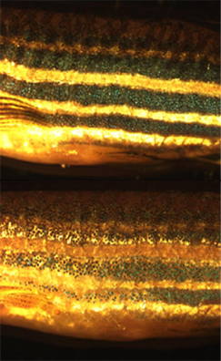
During embryonic development, chromatophores are one of a number of cell types generated in the neural crest. These cells have the ability to migrate long distances, allowing chromatophores to populate the entire surface of the body. Leaving the neural crest in waves, chromatophores take a dorsolateral route through the dermis, entering the ectoderm through small holes in the basal lamina. When and how multipotent chromatophore precursor cells differentiate into their different subtypes is an area of intense research. It is known in zebrafish embryos, for example, that by 3 days post fertilization all three pigment cells classes found in the fish - melanophores, xanthophores and iridophores - are already present.[23] Zebrafish larvae are also used in the to study how chromatophores undergo controlled proliferation and migration during embryogenesis to accurately generate the regular horizontal striped pattern in seen in adult fish.[24] These studies are seen as useful models for understanding colour patterning in the evolutionary developmental biology field.

Cephalopod chromatophores
Most notable in brightly coloured squid, cuttlefish and octopuses, cephalopods have complex multicellular 'organs' which they use to change colour rapidly. Each chromotophore unit is composed of a single chromatophore cell and numerous muscle, nerve, glial and sheath cells.[25] Inside the chromatophore cell, pigment granules are inclosed in an elastic sac, called the cytoelastic sacculus. To change colour the animal distorts the sacculus form or size by muscular contraction, thus changing its translucency, reflectivity or opacity. This differs from the mechanism used in fish, amphibians and reptiles, in that the shape of the sacculus is being changed rather than a translocation of pigment vesicles within the cell.
Octopuses operate chromatophores in complex, multi-cellular chromatic displays, resulting in a spectacular variety of colour schemes. Often repetitive waves of colour changes are observed as the neurons controlling the muscular contractions are patterned in the basal and peduncle lobes of the brain.[26] Like chameleons, cephalopods use physiological colour change for social interaction between individuals and as warning cues to other animals. They are also among the most adept at background adaptation, having the ability to match both the colour and the texture of their local environment with remarkable accuracy.
Notes
- ^ a b Bagnara JT. Cytology and cytophysiology of non-melanophore pigment cells. Int Rev Cytol. 1966; 20:173-205. Cite error: The named reference "Cytology" was defined multiple times with different content (see the help page).
- ^ Matsumoto J. Studies on fine structure and cytochemical properties of erythrophores in swordtail, Xiphophorus helleri. J Cell Biol. 1965; 27:493-504.
- ^ Taylor JD. The effects of intermedin on the ultrastructure of amphibian iridophores. Gen Comp Endocrinol. 1969; 12:405-16.
- ^ Morrison RL. A transmission electron microscopic (TEM) method for determining structural colors reflected by lizard iridophores. Pigment Cell Res. 1995; 8:28-36.
- ^ a b c d Fujii R. The regulation of motile activity in fish chromatophores. Pigment Cell Res. 2000; 13:300-19. Cite error: The named reference "fish" was defined multiple times with different content (see the help page).
- ^ Ito S & Wakamatsu K. Quantitative analysis of eumelanin and pheomelanin in humans, mice, and other animals: a comparative review. Pigment Cell Res. 2003; 16:523-31.
- ^ Bagnara JT et al. Color changes, unusual melanosomes, and a new pigment from leaf frogs. Science. 1973; 182:1034-5.
- ^ Schwalm PA et al. Infrared reflectance in leaf-sitting neotropical frogs. Science. 1977; 196:1225-7.
- ^ Bagnara JT et al. The dermal chromatophore unit. J Cell Biol. 1968; 38:67-79.
- ^ Palazzo RE et al. Rearrangements of pterinosomes and cytoskeleton accompanying pigment dispersion in goldfish xanthophores. Cell Motil Cytoskeleton. 1989; 13:9-20.
- ^ Porras MG et al. Corazonin promotes tegumentary pigment migration in the crayfish Procambarus clarkii. Peptides. 2003; 24:1581-9.
- ^ Deacon SW et al. Dynactin is required for bidirectional organelle transport. J Cell Biol. 2003; 160:297-301.
- ^ Aspengren S et al. Noradrenaline- and melatonin-mediated regulation of pigment aggregation in fish melanophores. Pigment Cell Res. 2003; 16:59-64.
- ^ a b Logan DW et al. Regulation of pigmentation in zebrafish melanophores. Pigment Cell Res. 2006; 19:206-13.
- ^ Logan DW et al. Sequence characterization of teleost fish melanocortin receptors. Ann N Y Acad Sci. 2003; 994:319-30.
- ^ Sugden D et al. Melatonin, melatonin receptors and melanophores: a moving story. Pigment Cell Res. 2004; 17:454-60.
- ^ Logan DW et al. The structure and evolution of the melanocortin and MCH receptors in fish and mammals. Genomics. 2003; 81:184-91.
- ^ Valverde P et al. Variants of the melanocyte-stimulating hormone receptor gene are associated with red hair and fair skin in humans. Nat Genet. 1995; 11:328-30.
- ^ Snider J et al. Intracellular actin-based transport: how far you go depends on how often you switch. Proc Natl Acad Sci USA. 2004; 101:13204-9.
- ^ Rodionov VI et al. Functional coordination of microtubule-based and actin-based motility in melanophores. Curr Biol. 1998; 8:165-8.
- ^ Rodionov VI et al. Switching between microtubule- and actin-based transport systems in melanophores is controlled by cAMP levels. Curr Biol. 2003; 13:1837-47.
- ^ Neuhauss SC. Behavioral genetic approaches to visual system development and function in zebrafish. J Neurobiol. 2003; 54:148-60.
- ^ Kelsh RN et al. Genetic analysis of melanophore development in zebrafish embryos. Dev Biol. 2000; 225:277-93.
- ^ Kelsh RN. Genetics and evolution of pigment patterns in fish. Pigment Cell Res. 2004; 17:326-36.
- ^ Cloney RA. & Florey E. Ultrastructure of cephalopod chromatophore organs. Zeits. für Zellforsch. 1968; 89:250-280.
- ^ Demski LS. Chromatophore systems in teleosts and cephalopods: a levels oriented analysis of convergent systems. Brain Behav Evol. 1992; 40:141-56.
