
Hypospadias
This article has multiple issues. Please help improve it or discuss these issues on the talk page. (Learn how and when to remove these template messages)
|
| Hypospadias | |
|---|---|
| Specialty | Medical genetics, urology |
Hypospadias /haɪpoʊˈspeɪdɪəs/[1][2] is a birth defect of the urethra in males and females that involves an abnormally placed urinary hole (the opening, or male external urethral orifice). Instead of opening at the tip of the glans of the penis, a hypospadic urethra opens anywhere along a line (the urethral groove) running from the tip along the underside (ventral aspect) of the shaft to the junction of the penis and scrotum or perineum, as is the case in pseudovaginal perineoscrotal hypospadias. A distal hypospadias may be suspected in an uncircumcised boy from an abnormally formed foreskin and downward tilt of the glans.
Hypospadia is a frequent birth difference in sex development consisting in fissure of the posterior (lower) wall of the urethra. Hypospadia is characterized by shortening of the urethra and ectopia of the external urethral opening. This difference in sex development (DSD), often occurs in women but is rarely diagnosed because physicians are not well-informed about female hypospadia. Classification of anatomic variants of female hypospadias includes low vaginal ectopia of the external urethral opening; high vaginal ectopy of the external opening of the urethra; urovaginal (vesicovaginal) fusion of the neck of the urinary bladder with vagina accompanied with enuresis; urogenital sinus in females (ectopia of the external urethral opening in the urogenital sinus); any of the above variants of female hypospadias in combination with false or true hermaphroditism. Hypospadias, urogenital sinus and hermaphroditism are three anomalies of human urogenital system which combine rather frequently.[3]
The urethral meatus opens on the underside of the glans penis in about 50–75% of cases; these are categorized as first degree hypospadias. Second degree (when the urethra opens on the shaft), and third degree (when the urethra opens on the perineum) occur in up to 20 and 30% of cases respectively. The more severe degrees are more likely to be associated with chordee, in which the phallus is incompletely separated from the perineum or is still tethered downwards by connective tissue, or with undescended testicles (cryptorchidism).[citation needed]
Types[4]
- Glandular hypospadias – most common variety
- Coronal hypospadias – meatus is placed at junction ofe shaft
- Perineal hypospadias – scrotum is split and urethra opens between its two halves
Signs and symptoms
Hypospadias is a male birth defect in which the opening of the tube that carries urine from the body (urethra) develops abnormally, usually on the underside of the penis. The opening can occur anywhere from just below the end of the penis to the scrotum. A form of hypospadias in which the genitals are abnormally positioned can also develop in males. Hypospadias are usually associated with a forward curvature of the penis, often referred to as chordee.[5]
Associated birth defects
Mild hypospadias most often occurs as an isolated birth defect without detectable abnormality of the remainder of the reproductive or endocrine system. However, a minority of infants, especially those with more severe degrees of hypospadias will have additional structural anomalies of the genitourinary tract. Up to 10% of boys with hypospadias have at least one undescended testicle, and a similar number have an inguinal hernia. An enlarged prostatic utricle is common when the hypospadias is severe (scrotal or perineal), and can predispose to urinary tract infections, pseudo-incontinence, or even stone formation.[citation needed]
Epispadias
A much rarer and unrelated type of urethral malformation is an epispadias. This is not a problem of the urethral groove or meatus, but a failure of abdominal and pelvic fusion in the first months of embryogenesis. An isolated opening of the dorsal ("top") side of the penis is rare, and most of these children have much more severe defects, involving a small and bifid phallus with bladder exstrophy or more severely, cloacal exstrophy involving the entire perineum. The cause of this defect of early embryogenesis is unknown but does not involve androgens.[5]
-
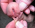 Example of penis with hypospadias
 -
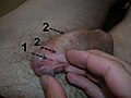 Penis with hypospadias (1) and two fistulae (2)


Causes
In most cases, the cause of this birth defect is not fully understood. Treatment with hormones such as progesterone during pregnancy may increase the risk of hypospadias.[6] Certain hormonal fluctuations, such as failure of the fetal testes to produce enough testosterone or the failure of the body to respond to testosterone, increase the risk of hypospadias and other genetic problems. Sometimes hypospadias is inherited.
There may also be an increased risk of hypospadias in infant males born to women of an advanced age or those who used in vitro fertilization (IVF) to conceive. The connection to IVF may be due to the mother's exposure to progesterone, a natural hormone, or to progestin, a synthetic form of progesterone, administered during the IVF process.[6][7]
Prenatal testosterone, converted in the genital skin to dihydrotestosterone, causes migration of skin fibroblasts to fully enclose the urethral groove in fetal males, normally resulting in an enclosed penile urethra by the second trimester of pregnancy. Failure of adequate prenatal androgen effect is therefore thought to be involved in many cases, making severe hypospadias a very mild form of intersex (under-virilization of a genetic male). Since postnatal androgen deficiency can only be demonstrated in a minority of cases, it has been proposed that transient deficiency of testosterone can occur during critical periods of fetal genital development, due to elevation of anti-müllerian hormone or more subtle degrees of pituitary-gonadal dysfunction. More recently, abnormalities of transcription factors have been proposed.[citation needed]
In animals, several teratogenic drugs or chemicals can cause hypospadias by interfering with androgen action in the embryo. Speculation that environmental agents—endocrine disruptors—might be interfering with human hormone systems has not been proven. The agents that have caused hypospadias in a small number of boys have been maternal use of synthetic progestins and finasteride in the first two trimesters of pregnancy. In 2008, it was suggested that maternal use of diethylstilbestrol, a synthetic estrogen, resulted in a 20-fold increase in prevalence of hypospadias[8] although a follow-up study showed the risk, though present, to be much lesser.[9]
In a minority of cases a postnatal deficiency of, or reduced sensitivity to, androgens (testosterone and dihydrotestosterone) can be demonstrated. These are often associated with a chordee, and in severe cases a residual perineal urogenital opening and small phallus. This combination of birth defects is referred to as pseudovaginal perineoscrotal hypospadias and is part of the spectrum of ambiguous genitalia. Treatment with testosterone postnatally does not close the urethra.[citation needed]
Genetic factors are likely involved in at least some cases, as there is about a 7% familial recurrence risk. A 2010 Article[10][11] found a 2.5 times increase in the condition for boys with a specific genetic defect that was carried on the X (maternally contributed sex) chromosome.
Rare iatrogenic urethral injuries similar to hypospadias after procedures such as surgery, catheterization, or circumcision have been reported.[citation needed]
Treatment
First degree hypospadias are primarily a cosmetic defect and have little effect on function except for direction of the urinary stream. If uncorrected, a second or third degree hypospadias can make urination messy, necessitate that it be performed sitting, impair delivery of semen into the vagina (possibly creating problems with fertility), or interfere with erections. In developed countries, most hypospadias are surgically repaired in infancy. Surgical repair of first and second degree hypospadias is nearly always successful in one procedure, usually performed in the first year of life by a pediatric urologist or a plastic surgeon.
When the hypospadias is third degree, or there are associated birth defects such as chordee or cryptorchidism, the best management can be a more complicated decision. A karyotype and endocrine evaluation should be performed to detect intersex conditions or hormone deficiencies. If the penis is small, testosterone or human chorionic gonadotropin (hCG) injections may be given to enlarge it before surgery.[12]
Surgical repair of severe hypospadias may require multiple procedures and mucosal grafting. Preputial skin is often used for grafting and circumcision should be avoided before repair. In a minority of patients with severe hypospadias surgery produces unsatisfactory results, such as scarring, curvature, or formation of urethral fistulas, diverticula, or strictures. A fistula is an unwanted opening through the skin along the course of the urethra, and can result in urinary leakage or an abnormal stream. A diverticulum is an "outpocketing" of the lining of the urethra which interferes with urinary flow and may result in post-urination leakage. A stricture is a narrowing of the urethra severe enough to obstruct flow. Reduced complication rates even for third degree repair (e.g., fistula rates below 5%) have been reported in recent years from centers with the most experience, and surgical repair is now performed for the vast majority of infants with hypospadias.[citation needed]
Due to the difficulties and lower success rates of surgical repair of the most severe degrees of under virilization, some of these genetically male but severely undervirilized infants have been assigned and raised as girls, with feminizing surgical reconstruction. Opinion has shifted against this approach in the last decade because adult sexual function as a female has often been poor, and development of a male gender identity despite female sex assignment and rearing has occurred in some XY children after reassignment for a more severe type of genital birth defect, cloacal exstrophy.[citation needed]
Epidemiology
Hypospadias are among the most common birth defects of the male genitalia (second to cryptorchidism), but widely varying incidences have been reported from different countries, from as low as 1 in 4000 to as high as 1 in 125 boys.[13]
Due to variations in the reporting requirements of different national databases, data from such registries can not be used to accurately determine either incidence of hypospadias or geographical variations in its occurrences.[12]
The incidence of hypospadias around the world has been increasing in recent decades. In the United States, two surveillance studies reported that the incidence had increased from about 1 in 500 total births (1 in 250 boys) in the 1970s to 1 in 250 total births (1 in 125 boys) in the 1990s. Although a slight worldwide increase in hypospadias was reported in the 1980s, studies in different countries and regions have yielded conflicting results and some registries have reported decreases. [citation needed]
See also
- Pediatric urology
- Andrology
- Cryptorchidism
- Bladder exstrophy, cloacal exstrophy
- Perineal urethra, pseudovaginal perineoscrotal hypospadias
- Ambiguous genitalia, intersex, intersex surgery
- Androgen insensitivity syndrome
References
- ^ Entry "hypospadias" in Merriam-Webster Online Dictionary.
- ^ OED 2nd edition, 1989 as /hɪpəʊˈspeɪdɪəs/~/haɪpəʊˈspeɪdɪəs/.
- ^ http://www.ncbi.nlm.nih.gov/m/pubmed/17722617/
- ^ Bailey & Love's/25th/1362-1363
- ^ a b King, Sebastian; Beasley, Spencer (2012) [1st. Pub. 1986]. "Chapter 9.1:Surgical Conditions in Older Children". In South, Mike (ed.). Practical Paediatrics, Seventh Edition. Churchill Livingstone, Elsevier. pp. 266–267. ISBN 978-0-702-04292-8.
{{cite book}}: Unknown parameter|lastauthoramp=ignored (|name-list-style=suggested) (help) - ^ a b Silver, RI; Rodriguez, R; Chang, TS; Gearhart, JP (June 1999). "In vitro fertilization is associated with an increased risk of hypospadias". The Journal of urology. 161 (6): 1954–7. doi:10.1016/S0022-5347(05)68863-5. PMID 10332480.
- ^ "Hypospadias: Risk Factors". The Mayo Clinic.
- ^ Klip H, Verloop J, van Gool JD, Koster ME, Burger CW, van Leeuwen FE (2002). "Hypospadias in sons of women exposed to diethylstilbestrol in utero: a cohort study". Lancet. 359 (9312): 1102–7. doi:10.1016/S0140-6736(02)08152-7. PMID 11943257.
{{cite journal}}: CS1 maint: multiple names: authors list (link) - ^ Brouwers MM, Feitz WF, Roelofs LA, Kiemeney LA, de Gier RP, Roeleveld N (2006). "Hypospadias: a transgenerational effect of diethylstilbestrol?". Hum. Reprod. 21 (3): 666–9. doi:10.1093/humrep/dei398. PMID 16293648.
{{cite journal}}: CS1 maint: multiple names: authors list (link) - ^ Loes F M van der Zanden; et al. (2010). "Common variants in DGKK are strongly associated with risk of hypospadias". Nature Genetics. 43 (1): 48–50. doi:10.1038/ng.721. PMID 21113153. Retrieved 2010-11-29.
{{cite journal}}: Explicit use of et al. in:|author=(help) - ^ "Clue found to penis birth defect". BBC News. 2010-11-29. Retrieved 2010-11-29.
- ^ a b Snodgrass, Warren (2012). "Chapter 130: Hypospadias". In Wein, Allan (ed.). Campbell-Walsh Urology , Tenth Edition. Elsevier. pp. 3503–3536. ISBN 9781416069119.
- ^ Gatti, John M. "Epidemiology". Medscape Reference. Retrieved 22 January 2013.
- Austin PF, Siow Y, Fallat ME, Cain MP, Rink RC, Casale AJ (2002). "The relationship between müllerian inhibiting substance and androgens in boys with hypospadias". J. Urol. 168 (4 Pt 2): 1784–8, discussion 1788. doi:10.1016/S0022-5347(05)64413-8. PMID 12352359.
{{cite journal}}: CS1 maint: multiple names: authors list (link) - Patel RP, Shukla AR, Snyder HM (2004). "The island tube and island onlay hypospadias repairs offer excellent long-term outcomes: a 14-year followup". J. Urol. 172 (4 Pt 2): 1717–9, discussion 1719. doi:10.1097/01.ju.0000138903.20136.22. PMID 15371798.
{{cite journal}}: CS1 maint: multiple names: authors list (link) - Retik AB, Atala A (2002). "Complications of hypospadias repair". Urol. Clin. North Am. 29 (2): 329–39. doi:10.1016/S0094-0143(02)00026-5. PMID 12371224.
- Shukla AR, Patel RP, Canning DA (2004). "Hypospadias". Urol. Clin. North Am. 31 (3): 445–60, viii. doi:10.1016/j.ucl.2004.04.020. PMID 15313054.
{{cite journal}}: CS1 maint: multiple names: authors list (link) - "Hairspray is linked to common genital birth defect".
{{cite journal}}: Cite journal requires|journal=(help)
External links
- International Trends in Rates of Hypospadias and Cryptorchidism
- The Hypospadias and Epispadias Association, for families affected by these congenital penile differences.
- Review of scientific papers describing the physical and psychological implications and consequences of hypospadias.
- Uretheral graft technique (Camillo Il Grande)
- "Special issue on hypospadias". Indian Journal of Urology. 24 (2). 2008.
- Interactive animations on hypospadias, AboutKidsHealth.ca