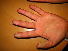
Desquamation
| Desquamation |
|---|
Desquamation (from Latin desquamare 'to scrape the scales off a fish'), also called skin peeling, is the shedding of the outermost membrane or layer of a tissue, such as the skin.
Skin


Normal, nonpathologic desquamation of the skin occurs when keratinocytes, after moving typically over about 14 days, are individually shed unnoticeably.[1] In pathologic desquamation, such as that seen in X-linked ichthyosis, the stratum corneum becomes thicker (hyperkeratosis), imparting a "dry" or scaly appearance to the skin, and instead of detaching as single cells, corneocytes are shed in clusters, forming visible scales.[1] Desquamation of the epidermis may result from disease or injury of the skin. For example, once the rash of measles fades, there is desquamation. Skin peeling typically follows healing of a first degree burn or sunburn. Toxic shock syndrome, a potentially fatal immune system reaction to a bacterial infection such as Staphylococcus aureus,[2] can cause severe desquamation; so can mercury poisoning. Other serious skin diseases involving extreme desquamation include Stevens–Johnson syndrome and toxic epidermal necrolysis (TEN).[3] Radiation can cause dry or moist desquamation.[4]
Eyes
Eye tissues including the conjunctiva and cornea may undergo pathological desquamation in diseases such as dry eye syndrome.[5] The anatomy of the human eye makes desquamation of the lens impossible.[6]
See also
- desquamative gingivitis
- Pityriasis—flaking of the skin
- Spalling
- Sunburn
- Moist desquamation
References
- ^ a b Jackson, Simon M.; Williams, Mary L.; Feingold, Kenneth R.; Elias, Peter M. (1993). "Pathobiology of the Stratum Corneum". The Western Journal of Medicine. 158 (3): 279–85. PMC 1311754. PMID 8460510.
- ^ Dinges, MM; Orwin, PM; Schlievert, PM (January 2000). "Exotoxins of Staphylococcus aureus". Clinical Microbiology Reviews. 13 (1): 16–34, table of contents. doi:10.1128/cmr.13.1.16-34.2000. PMC 88931. PMID 10627489.
- ^ Parillo, Steven J; Parillo, Catherine V. (2010-05-25). "Stevens-Johnson Syndrome". eMedicine. Medcape. Retrieved 2010-09-06.
- ^ Centers for Disease Control and Prevention (2005-06-30). "Cutaneous Radiation Injury". CDC. Retrieved 2011-05-15.
- ^ Gilbard, Jeffrey P. (November 1, 2003). "Dry Eye: Natural History, Diagnosis and Treatment". Wolters Kluwer Pharma Solutions. Retrieved February 3, 2012.
- ^ Lynnerup, Niels; Kjeldsen, Henrik; Heegaard, Steffen; Jacobsen, Christina; Heinemeier, Jan (2008). Gazit, Ehud (ed.). "Radiocarbon Dating of the Human Eye Lens Crystallines Reveal Proteins without Carbon Turnover throughout Life". PLoS ONE. 3 (1): e1529. doi:10.1371/journal.pone.0001529. PMC 2211393. PMID 18231610.
{{cite journal}}: CS1 maint: unflagged free DOI (link)
