External anal sphincter
| Sphincter ani externus muscle | |
|---|---|
 | |
 Coronal section through the anal canal. B. Cavity of urinary bladder V.D. Ductus deferens. S.V. Seminal vesicle. R. Second part of rectum. A.C. Anal canal. L.A. Levator ani. I.S. Sphincter ani internus. E.S. Sphincter ani externus. | |
| Details | |
| Nerve | Branch from the fourth sacral and contributions from the inferior hemorrhoidal branch of the pudendal nerve |
| Actions | Keep the anal canal and orifice closed |
| Identifiers | |
| Latin | Musculus sphincter ani externus |
| TA98 | A04.5.04.012 |
| TA2 | 2426 |
| FMA | 21930 |
| Anatomical terms of muscle | |
The external anal sphincter (or sphincter ani externus ) is a flat plane of muscular fibers, elliptical in shape and intimately adherent to the skin surrounding the margin of the anus.
Anatomy
The external anal sphincter measures about 8 to 10 cm in length, from its anterior to its posterior extremity, and is about 2.5 cm opposite the anus, when defecation occurs the sphincter muscle retracts.
It consists of two strata, superficial and deep.
- The superficial, constituting the main portion of the muscle, arises from a narrow tendinous band, the anococcygeal raphe, which stretches from the tip of the coccyx to the posterior margin of the anus; it forms two flattened planes of muscular tissue, which encircle the anus and meet in front to be inserted into the central tendinous point of the perineum, joining with the superficial transverse perineal muscle, the levator ani, and the bulbospongiosus muscle also known as the bulbocavernosus.
- The deeper portion forms a complete sphincter to the anal canal. Its fibers surround the canal, closely applied to the internal anal sphincter, and in front blend with the other muscles at the central point of the perineum.
In a considerable proportion of cases the fibers decussate in front of the anus, and are continuous with the superficial transverse perineal muscle.
Posteriorly, they are not attached to the coccyx, but are continuous with those of the opposite side behind the anal canal.
The upper edge of the muscle is ill-defined, since fibers are given off from it to join the levator ani.
Actions
The action of this muscle is peculiar.
(1) It is, like other muscles, always in a state of tonic contraction, and having no antagonistic muscle it keeps the anal canal and orifice shut.
(2) It can be put into a condition of greater contraction under the influence of the will, so as more firmly to occlude the anal aperture, in expiratory efforts unconnected with defecation.
(3) Taking its fixed point at the coccyx, it helps to fix the central point of the perineum, so that the bulbospongiosus muscle may act from this fixed point.
Additional images
-
Intestines
-
Schematic demonstrating the anatomy of hemorrhoids.
-
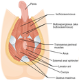
Muscles of male perineum.
-
Muscles of the female perineum.
-
Saggital (Vertical) section of bladder, penis, and urethra.
See also
References
![]() This article incorporates text in the public domain from page 425 of the 20th edition of Gray's Anatomy (1918)
This article incorporates text in the public domain from page 425 of the 20th edition of Gray's Anatomy (1918)
External links
- Anatomy photo:42:13-0100 at the SUNY Downstate Medical Center - "The Male Perineum and the Penis: The External Anal Sphincter"
- perineum at The Anatomy Lesson by Wesley Norman (Georgetown University) (analtriangle3)
- pelvis at The Anatomy Lesson by Wesley Norman (Georgetown University) (rectum)


