User:Ulflund/sandbox
To do
[edit]- Expand X-ray optics
- Expand X-ray_detector#Scintillators
- Birefringence
- Clean up drift velocity
- Go through Wikipedia:WikiProject_Physics/Missing_physics_topics
- Read [[1]]
- Improve Image noise and Image articles
- Improve X-ray
- Create Beam hardening article
- Create X-ray detector article
- Generalize field of view article to include microscopy definition
- Clean up spatial frequency article
- Refractive index
- X-rays (new section)
- Phase velocity
- Gases
Bulk
[edit]Aluminium metal has an appearance ranging from silvery to dull gray, depending on the surface roughness. A fresh film of aluminium serves as a good reflector (approximately 92%) of visible light and an excellent reflector (as much as 98%) of medium and far infrared radiation.
The density of aluminium is 2.70 g/cm3, about 1/3 that of steel, much lower than other commonly encountered metals, making aluminium parts easily identifiable through their lightness.[1][a] Aluminium is not as strong or stiff as steel, but the low density makes up for this in the aerospace industry and for many other applications where light weight is crucial.
Pure aluminum is quite soft and lacking in strength. In most applications various aluminium alloys are used instead because of their higher strength and hardness. The yield strength of pure aluminium is 7–11 MPa, while aluminium alloys have yield strengths ranging from 200 MPa to 600 MPa.[2]
Aluminium is ductile, and malleable allowing it to be easily drawn and extruded. It is also easily machined, and the low melting temperature of 660 °C allows for easy casting.
Aluminium is a good thermal and electrical conductor, having 59% the conductivity of copper, both thermal and electrical, while having only 30% of copper's density. Aluminium is capable of superconductivity, with a superconducting critical temperature of 1.2 kelvin and a critical magnetic field of about 100 gauss (10 milliteslas).[3] It is paramagnetic and thus essentially unaffected by static magnetic fields. The high electrical conductivity, however, means that it is strongly affected by changing magnetic field through the induction of eddy currents.
X-ray optics
[edit]- Focusing optics
- Mirrors
- Compound refractive lenses
- Zone plates
- Polycapillary lenses
- Monocapillary lenses
- Filters
- Monocromators
- Gratings
X-ray mirrors
[edit]Curved mirrors are often used to focus x-rays, both at synchrotron beam lines, in compact setups and for x-ray astronomy. To achieve decent reflectivity the geometry is typically such that the x-rays are reflected at grazing incidence angles, i.e. with propagation directions almost parallell to the mirror surface. At low enough glancing angles x-rays get totally externally reflected resulting in close to 100% reflectivity. This occurs below the critical angle, which depend on the mirror material and the x-ray wavelength, but is often below 1°.
At larger angles decent reflectivity can be achieved by coating the mirror surface in a multilayer optical coating with many, often hundreds, of alternating thin layers of a dense material such as tungsten or gold and a light material such as coal or silicon. If the layer thicknesses, which are typically one to a few nanometer, are just right, the reflections from all the individual layers will interfere constructively and thus amplify the reflected beam. For hard x-rays grazing angles can be up to a few degrees, but for soft x-rays, which have longer wavelengths and thus need thicker layers, mirrors can be made even for normal incidence reflections.[4]
Other focusing optics
[edit]There are also a few other less commonly used techniques for focusing x-rays. Microstructed optical arrays are made by etching a large number of small channels into a substrate. X-rays are focused through total external reflection on the inside of these channels. There can be one substrate with parallell channels providing a one-to-one imaging[5] or two consecutive array components in order to reduce comatic aberration. Microstructed optical arrays are used in applications which require x-ray focal spots in the order of few micrometers or below, such as radiobiology of individual cells.
X-rays
[edit]The complex refractive index for x-rays is for all materials close to 1 with the real pars slightly smaller than 1. For this reason it is usually written as , where the refractive index decrement is responsible for refraction while the imaginary part is responsible for attenuation of the x-rays.
X-ray
[edit]- Reorder sections, move exposure down
- separate detectors into imaging detectors and single pixel detectors, possibly as a new article
soft x-ray limit
[edit]What different sources say about where the limit between soft and hard x-rays is.
- 10 keV, [2] Hard x-rays are typically those with energies greater than around 10 keV.
- 6-8 keV, [3] Figure in presentation shows a soft limit between 6 and 8 keV.
- 4.1 keV, [4] The picture above was taken with a camera that sees light with wavelengths between about 0.3 and 4.5 nanometers, the so-called "Soft X-rays."
- 3.1 keV, [5] Table giving the limit at 4 Å.
- 12 keV, [6] Wolfram alpha limit at 3e9 GHz
- 5-10 keV, Peter Skoglund licentiate thesis p. 1.
- 5-6 keV, Michael Bertilson Doctoral thesis
- 6 keV, [7] in the USSR, the wavelength dividing hard X rays from soft X rays is usually taken as 2 A.
Source table
[edit]| Anode material |
Atomic number |
Photon energy [keV] | Wavelength [nm] | ||
|---|---|---|---|---|---|
| Kα1 | Kβ1 | Kα1 | Kβ1 | ||
| W | 74 | 59.3 | 67.2 | 0.0209 | 0.0184 |
| Mo | 42 | 17.5 | 19.6 | 0.0709 | 0.0632 |
| Cu | 29 | 8.05 | 8.91 | 0.157 | 0.139 |
| Ag | 47 | 22.2 | 24.9 | 0.0559 | 0.0497 |
| Ga | 31 | 9.25 | 10.26 | 0.134 | 0.121 |
| In | 49 | 24.2 | 27.3 | 0.0512 | 0.455 |
X-ray detector
[edit]X-ray detectors are devices used to measure the flux, spatial distribution, spectrum or other properties of X-rays. They vary in shape and function depending on their purpose. Some common principles used to detect X-rays include the ionization of gas, the conversion to visible light in a scintillator and the production of electron-hole pairs in a semiconductor detector. Imaging detectors such as those used for radiography where originally based on photographic plates and later photographic film but are now mostly replaced by various digital detector types such as image plates or flat panel detectors.
For radiation protection direct exposure hazard is often evaluated using ionization chambers, while dosimeters are used to measure the radiation dose a person has been exposed to. X-ray spectra can be measured either by energy dispersive or wavelength dispersive spectrometers.
X-ray detectors can be either photon counting or integrating. Photon-counting detectors measure each individual x-ray photon separately, while integrating detectors measure the total amount of energy deposited in the active region of the detector. Photon-counting detectors are normally more sensitive since they do not suffer from thermal and readout noise in the same way. An other advantage is that they can be set to count only photons in a certain energy range, or even messure the energy of each absorbed photon. Integrating detectors are normally simpler and can handle much higher photon fluxes.
The purpose can vary from radiation protection to medical imaging or material analysis. Detectors can be divided into spatially resolving detectors used for imaging and single-pixel detectors used for e.g. dosimetry and spectral analysis. Another distinction is between photon-counting and integrating detectors.
X-ray detectors are based on various methods. The most commonly known methods are photographic plates, photographic film in cassettes, and rare earth screens. Regardless of what is "catching" the image, they are all categorized as "Image Receptors" (IR).
Most X-ray detectors utilize the ionising capability of X-rays which means that they can often be used to measure also other types of ionizing radiation.
Gas detectors
[edit]
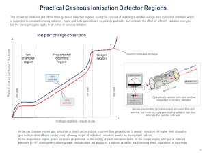
X-rays going through a gas will ionize it, producing positive ions and free electrons. An incoming photon will create a number of such ion pairs proportional to its energy. If there is an electric field in the gas chamber ions and electrons will move in different directions and thereby cause a detectable current. The behaviour of the gas will depend on the applied voltage and the geometry of the chamber. This gives rise to a few different types of gas detectors described below.
Ionization chambers use a relatively low electric field of about 100 V/cm to extract all ions and electrons before they recombine.[8] This gives a steady current proportional to the dose rate the gas is exposed to. Ion chambers are widely used as hand held radiation survey meters to check radiation dose levels.
Proportional counters use a geometry with a thin positively charged anode wire in the center of a cylindrical chamber. Most of the gas volume will act as an ionization chamber, but in the region closest to the wire the electric field is high enough to make the electrons ionize gas molecules. This will create an avalanche effect greatly increasing the output signal. Since every electron cause an avalanch of approximately the same size the collected charge is proportional to the number of ion pairs created by the absorbed x-ray. This makes it possible to measure the energy of each incoming photon.
Geiger–Müller counters use an even higher electric field so that UV-photons are created. These start new avalanches eventually resulting in a total ionization of the gas around the anode wire. This makes the signal very strong, but causes a dead time after each event and makes it impossible to measure the X-ray energies.
Gas detectors can be made spatially resolving by having many crossed wires in a wire chamber.
Scintillator-based detectors
[edit]Scintillators are materials that absorb x-rays and emmit visible light. They can be used to measure the total x-ray flux or to count individual photons.
In a scintillation counter a scintillator is optically coupled to a light-sensitive device such as a photomultiplier tube or a silicon PIN photodiode.[9]
X-ray image intensifier http://radiographics.rsna.org/content/20/5/1471.full
Scintillator coupled to CCD or CMOS detector
Medical imaging detectors
[edit]- photographic plates
- image plates
- flat panel detectors
- rare earth screen?
Semiconductor detectors
[edit]- Semiconductor detectors
- Direct detection CCDs
- Pilatus (detector)
Superconducting detectors
[edit]Astronomical x-ray detectors
[edit]- Microchannel plate detectors
- X-ray calorimeters [8]
Dosimeters
[edit]Dosimeters are devices worn by the user which measure the equivalent dose that the user is receiving.
- Quartz fiber dosimeter
- Film badge dosimeter
- Thermoluminescent dosimeter
- Solid state (MOSFET or silicon diode) dosimeter
sources
[edit]http://imagine.gsfc.nasa.gov/docs/science/how_l1/xray_detectors.html
X-ray phase contrast
[edit]There are four main techniques for x-ray phase-contrast imaging, all utilizing different principles to convert phase variations in the x-rays emerging from the object, into intensity variations at an x-ray detector.[10][11] Propagation-based phase contrast[12] uses free-space propagation to get edge enhancement, talbot interferometry[11] uses a set of diffraction gratings to measure the derivative of the phase, refraction-enhanced imaging[13] uses an analyzer crystal also for differential measurement, and x-ray interferometry[14] uses a crystal interferometer to measure the phase directly. The advantage of these methods compared to normal absorption-contrast x-ray imaging is the higher contrast making it possible to see smaller details. One disadvantage is that these methods require more sophisticated equipment, such as synchrotron or microfocus x-ray sources, x-ray optics and high resolution x-ray detectors. The high demands on equipment is mainly due to the small variations in refractive index for x-rays. The refractive index is normally smaller than 1 with a difference from 1 between 10−7 and 10−6.
All of these methods produce images that can be used to calculate the projections (integrals) of the refractive index in the imaging direction. For propagation-based phase contrast there are phase-retrieval algorithms, for talbot interferometry and refraction-enhanced imaging the image is integrated in the proper direction, and for x-ray interferometry phase unwrapping is performed. For this reason they are well suited for tomography, i.e. reconstruction of a 3D-map of the refractive index of the object from many images at slightly different angles. For x-ray radiation the difference from 1 of the refractive index is essentially proportional to the density of the material.
Propagation-based phase contrast
[edit]Propagation-based phase contrast, in-line phase contrast, in-line holography or diffraction-enhanced imaging is the simplest method of achieving x-ray phase contrast imaging. ...
Talbot interferometry
[edit]Refraction-enhanced imaging
[edit]X-ray interferometry
[edit]- Lead
- As it is now.
- Definition
- Containing most of what is now in Work (physics)#Mathematical calculation. I'm not sure about the title for the section, but I don't like Mathematical calculation.
- Zero work
- A combination of the paragraph in Work (physics)#Mathematical calculation on zero work and the section Work (physics)#Zero work.
- Torque and roation
- With the same content as Work (physics)#Torque and rotation, but also including the case where torque and rotation are not parallell.
- Units
- same as current section Work (physics)#Units, but with the paragraph about heat removed.
- Work-energy theorem
- Same content as Work (physics)#Work and kinetic energy but with a correct proof also for the simple case (not requireing ).
- Power
- Same content as in Work (physics)#Moving objects and power but with the integrals and derivatives written out. Maybe this should instead be merged into the Definition section.
- Frame of reference
- Keep as is. Maybe write that the work-energy theorem only works in an inertial frame of reference unless fictitious forces are included.
The total work done by all forces on a rigid body is equal to that body's change in kinetic energy. This is called the work-energy theorem and ....
This theorem is particularly simple to prove for a constant force F acting on a point mass m moving parallell to the force. The mass will during a time t change its speed from to , where is the acceleration given by Newton's second law . This can be rewritten as
- .
The distance traveled is the average speed times the time, which for a constant acceleration is given by
- .
Using these equations we can expand the expression for work as
- .
For a point mass the kinetic energy is given by
- ,
which we can identify in the previous equation to get
- .
Total internal reflection
[edit][File:Réflexion total.svg|thumb|right|upright|The larger the angle to the normal, the smaller is the fraction of light transmitted rather than reflected, until the angle at which total internal reflection occurs. (The color of the rays is to help distinguish the rays, and is not meant to indicate any color dependence.)]]

In physics, total internal reflection is a phenomenon where a wave reaching a medium boundary is fully reflected. This occurs when the second medium has a lower refractive index than the first one and the incidence angle from the normal of the surface is larger than the critical angle for the pair of materials. This is particularly common as an optical phenomenon, where light waves are involved, but it occurs with many types of waves, such as electromagnetic waves in general or sound waves.
When a wave crosses a boundary between different materials with different kinds of refractive indices, the wave will be partially refracted at the boundary surface, and partially reflected. However, if the angle of incidence is greater (i.e. the direction of propagation or ray is closer to being parallel to the boundary) than the critical angle – the angle of incidence at which light is refracted such that it travels along the boundary – then the wave will not cross the boundary and instead be totally reflected back internally. This can only occur when the wave in a medium with a higher refractive index (n1) hits its surface that's in contact with a medium of lower refractive index (n2). For example, it will occur with light hitting air from glass, but not when hitting glass from air.

Refraction todo
[edit]General explanation
[edit]continuous case
Light
[edit]Add subsections:
Lenses
[edit]Birefringence
[edit]Sound
[edit]In air, Seismic refraction
Water waves
[edit]bend towards shores
References
[edit]- ^ Lide 2004, p. 4-3.
- ^ Polmear, I.J. (1995). Light Alloys: Metallurgy of the Light Metals (3 ed.). Butterworth-Heinemann. ISBN 978-0-340-63207-9.
- ^ Cochran, J.F.; Mapother, D.E. (1958). "Superconducting Transition in Aluminum". Physical Review. 111 (1): 132–142. Bibcode:1958PhRv..111..132C. doi:10.1103/PhysRev.111.132.
- ^ "Curved mirror optics". x-ray-optics.de. Retrieved 2016-12-17.
- ^ "Multiple flat mirrors". x-ray-optics.de. Retrieved 2016-12-14.
- ^ "X-ray Transition Energies". NIST Physical Measurement Laboratory. 2011-12-09. Retrieved 2013-03-10.
- ^ "X-Ray Data Booklet Section 1.2 X-ray emission energies". Center for X-ray Optics and Advanced Light Source, Lawrence Berkeley National Laboratory. 2009-10-01. Retrieved 2013-03-12.
- ^ Albert C. Thompson. X-Ray Data Booklet, Section 4-5 X-ray detectors (PDF).
- ^ "Scintillation counter". The free dictionary by Farlex.
{{cite web}}: Unknown parameter|retrieved=ignored (|access-date=suggested) (help) - ^ Fitzgerald, Richard (2000). "Phase-sensitive x-ray imaging". Physics Today. 53 (7): 23. doi:10.1063/1.1292471.
- ^ a b David, C, Nohammer, B, Solak, H H, & Ziegler E (2002). "Differential x-ray phase contrast imaging using a shearing interferometer". Applied Physics Letters. 81 (17): 3287–3289. doi:10.1063/1.1516611.
{{cite journal}}: CS1 maint: multiple names: authors list (link) - ^ Wilkins, S W, Gureyev, T E, Gao, D, Pogany, A & Stevenson, A W (1996). "Phase-contrast imaging using polychromatic hard X-rays". Nature. 384: 335–338. doi:10.1038/384335a0.
{{cite journal}}: CS1 maint: multiple names: authors list (link) - ^ Davis, T J, Gao, D, Gureyev, T E, Stevenson, A W & Wilkins, S W (1995). "Phase-contrast imaging of weakly absorbing materials using hard X-rays". Nature. 373: 595–598. doi:10.1038/373595a0.
{{cite journal}}: CS1 maint: multiple names: authors list (link) - ^ Momose, A, Takeda, T, Itai, Y & Hirano, K (1996). "Phase-contrast X-ray computed tomography for observing biological soft tissues". Nature Medicine. 2: 473–475. doi:10.1038/nm0496-473.
{{cite journal}}: CS1 maint: multiple names: authors list (link)
Cite error: There are <ref group=lower-alpha> tags or {{efn}} templates on this page, but the references will not show without a {{reflist|group=lower-alpha}} template or {{notelist}} template (see the help page).


