Median nerve: Difference between revisions
| Line 30: | Line 30: | ||
===Course and Branches in the forearm=== |
===Course and Branches in the forearm=== |
||
The median nerves arises from the cubital fossa and passes between the two heads of [[pronator teres]]. It then travels between [[flexor digitorum superficialis]] and [[flexor digitorum profundus]] before emerging between [[flexor digitorum superficialis]] and [[flexor carpi radialis]] |
The median nerves arises from the cubital fossa and passes between the two heads of [[pronator teres]]giving branches to it. It then travels between [[flexor digitorum superficialis]] and [[flexor digitorum profundus]] before emerging between the tendons of the [[flexor digitorum superficialis]] and [[flexor carpi radialis]]near the wrist and deep to the tendon of the palamaris longus |
||
The '''unbranched portion''' of the median nerve (which arises from the cubital fossa) innervates muscles of superficial and intermediate groups of the anterior compartment except flexor carpi ulnaris |
|||
The median nerve does give off two branches as it courses through the forearm: |
The median nerve does give off two branches as it courses through the forearm: |
||
Revision as of 22:09, 30 September 2007
| Median nerve | |
|---|---|
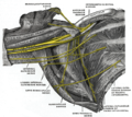
| File:Gray816.png Diagram from Gray's anatomy, depicting the peripheral nerves of the upper extremity, amongst others the median nerve | |
| Details | |
| From | Lateral cord, Medial cord |
| Innervates | Anterior compartment of the forearm (with two exceptions), Thenar eminence, Lumbricals |
| Identifiers | |
| Latin | nervus medianus |
| MeSH | D008475 |
| TA98 | A14.2.03.031 |
| TA2 | 6459 |
| FMA | 14385 |
| Anatomical terms of neuroanatomy | |
The median nerve is a nerve that runs down the arm and forearm. It is one of the five main nerves originating from the brachial plexus.
The median nerve is formed from parts of the medial and lateral cords of the brachial plexus, and continues down the arm to enter the forearm with the brachial artery.
The median nerve is the only nerve that passes through the carpal tunnel, where it may be compressed to cause carpal tunnel syndrome.
Course
Course in the Upper Arm
After receiving inputs from both the lateral and medial cords of the brachial plexus, the median nerve courses with brachial artery on medial side of arm between biceps brachii and brachialis. At first lateral to the artery, it then crosses anteriorly to run medial to the artery in the distal arm and into the cubital fossa.
The median nerve gives off no branches in the upper arm.
Course and Branches in the forearm
The median nerves arises from the cubital fossa and passes between the two heads of pronator teresgiving branches to it. It then travels between flexor digitorum superficialis and flexor digitorum profundus before emerging between the tendons of the flexor digitorum superficialis and flexor carpi radialisnear the wrist and deep to the tendon of the palamaris longus
The median nerve does give off two branches as it courses through the forearm:
- The anterior interosseous branch courses with the anterior interosseous artery and innervates all the muscles of the deep group of the anterior compartment of the forearm except the medial (ulnar) half of flexor digitorum profundus. Its ends with its innervation of pronator quadratus.
- The palmar cutaneous branch of the median nerve arises at distal part of the forearm. It supplies sensory innervation to the lateral aspect of the palmar skin (but not the digits).
The palmar cutaneous branch of the median nerve, which supplies the lateral aspect of the palmar skin arises proximal to the flexor retinaculum and passes superficial to it so does not pass through the carpal tunnel.
Branches in the hand
The median nerve enters the hand through the carpal tunnel, deep to the flexor retinaculum along with the tendons of flexor digitorum superficialis, flexor digitorum profundus, and flexor pollicis longus.
From there it sends off several branches:
- 1. Recurrent branch to muscles of the thenar compartment
- 2. Digital cutaneous branches to common palmar digital branch and proper palmar digital branch of the median nerve which supply the:
- a) lateral (radial) three and a half digits on the palmar side
- b) index, middle and ring finger on dorsum of the hand
The median nerve supplies motor innervation to the first and second lumbricals.
Innervation
Upper Arm
No motor innervation.
Forearm
It innervates most of the flexors in the forearm except flexor carpi ulnaris and the medial two digits of flexor digitorum profundus, which are supplied by the ulnar nerve.
Unbranched, the median nerves supplies the following muscles.
Superior Group:
Intermediate Group:
The anterior interosseus branch supplies the following muscles...
Deep group:
- lateral (radial) half of FDP
- FPL
- pronator quadratus
Hand
In the hand, the median nerve supplies motor innervation to the 1st and 2nd lumbricals and the muscles of the thenar eminence of the hand by a recurrent thenar branch. The rest of the intrinsic muscles of the hand are supplied by the ulnar nerve.
The median nerve innervates the skin of the palmar side of the thumb, the index and middle finger, half the ring finger, and the nail bed of these fingers. The lateral part of the palm is supplied by the palmar cutaneous branch of the median nerve which leaves the nerve proximal to the wrist creases. This palmar cutaneous branch travels in a separate fascial groove adjacent to the flexor carpi radialis.
Injury
Injury of median nerve at different levels cause different syndromes. Injury of this nerve at a level above elbow joint results in loss of pronation and a decrease in flexion of the hand at the wrist joint. In the hand, thenar muscles are paralysed and atrophy in time. Opposition and flexion movements of thumb are lost, and thumb and index finger are arrested in adduction and hyperextension position. This appearance of the hand is collectively referred as ape hand deformity. In addition, in palmar side of the hand sensation of lateral part of hand, first three fingers and lateral half of the fourth finger and in dorsal side sensation of distal ⅓ portions of first three fingers and lateral half of distal ⅓ portion of fourth finger is lost.
Additional images
-
Nervous system
-
Cross-section through the middle of upper arm.
-
Cross-section through the middle of the forearm.
-
Transverse section across distal ends of radius and ulna.
-
Transverse section across the wrist and digits.
-
The brachial artery.
-
Ulnar and radial arteries. Deep view.
-
The right brachial plexus (infraclavicular portion) in the axillary fossa; viewed from below and in front.
-
Cutaneous nerves of right upper extremity. Anterior view.
-
Diagram of segmental distribution of the cutaneous nerves of the right upper extremity. Anterior view.
-
Diagram of segmental distribution of the cutaneous nerves of the right upper extremity. Posterior view.
-
Superficial palmar nerves.
-
Deep palmar nerves.
-
Front of right upper extremity, showing surface markings for bones, arteries, and nerves.
-
Brachial plexus
External links
- Median nerve at the Duke University Health System's Orthopedics program
- Median+Nerve at the U.S. National Library of Medicine Medical Subject Headings (MeSH)
- Hand kinesiology at the University of Kansas Medical Center
- Atlas image: hand_plexus at the University of Michigan Health System - "Axilla, dissection, anterior view"
![]() This article incorporates text in the public domain from page 938 of the 20th edition of Gray's Anatomy (1918)
This article incorporates text in the public domain from page 938 of the 20th edition of Gray's Anatomy (1918)


