Matrix-assisted laser desorption/ionization: Difference between revisions
m Undid revision 208049541 by 203.193.135.81 (talk) |
|||
| Line 58: | Line 58: | ||
| 10,600 || (Overberg 1991)<ref>Overberg, A.; Karas, M.; Hillenkamp, F., Matrix-assisted Laser Desorption of Large Biomolecules with a TEA-CO<sub>2</sub>-Laser. Rapid Commun. Mass Spectrom. 1991, 5, 128-131.</ref> |
| 10,600 || (Overberg 1991)<ref>Overberg, A.; Karas, M.; Hillenkamp, F., Matrix-assisted Laser Desorption of Large Biomolecules with a TEA-CO<sub>2</sub>-Laser. Rapid Commun. Mass Spectrom. 1991, 5, 128-131.</ref> |
||
|} |
|} |
||
The [[laser]] is fired at the crystals in the MALDI spot. The matrix absorbs the laser energy and it is thought that primarily the matrix is ionized by this event. The matrix is then thought to transfer part of its charge to the analyte molecules (e.g. protein), thus ionizing them while still protecting them from the disruptive energy of the laser. Ions observed after this process consist of a neutral molecule [M] and an added or removed ion. Together, they form a quasimolecular ion, for example [M+H]<sup>+</sup> in the case of an added proton, [M+Na]<sup>+</sup> in the case of an added [[sodium]] ion, or [M-H]<sup>-</sup> in the case of a removed proton. MALDI generally produces singly-charged ions, but multiply charged ions ([M+nH]<sup>n+</sup>) can also be observed, usually as a function of the matrix, the laser intensity and/or the voltage used. Note that these are all even-electron species. Ion signals of radical cations can be observed eg. in case of matrix molecules and other stable molecules. |
The [[laser]] is fired at the crystals in the MALDI spot. The matrix absorbs the laser energy and it is thought that primarily the matrix is ionized by this event. The matrix is then thought to transfer part of its charge to the analyte molecules (e.g. protein), thus ionizing them while still protecting them from the disruptive energy of the laser. Ions observed after this process consist of a neutral molecule [M] and an added or removed ion. Together, they form a quasimolecular ion, for example [M+H]<sup>+</sup> in the case of an added proton, [M+Na]<sup>+</sup> in the case of an added [[sodium]] ion, or [M-H]<sup>-</sup> in the case of a removed proton. MALDI generally produces singly-charged ions, but multiply charged ions ([M+nH]<sup>n+</sup>) can also be observed, usually as a function of the matrix, the laser intensity and/or the voltage used. Note that these are all even-electron species. Ion signals of radical cations can be observed eg. in case of matrix molecules and other stable molecules. |
||
Revision as of 05:29, 25 April 2008

Matrix-assisted laser desorption/ionization (MALDI) is a soft ionization technique used in mass spectrometry, allowing the analysis of biomolecules (biopolymers such as proteins, peptides and sugars) and large organic molecules (such as polymers, dendrimers and other macromolecules), which tend to be fragile and fragment when ionized by more conventional ionization methods. It is most similar in character to electrospray ionization both in relative softness and the ions produced (although it causes many fewer multiply charged ions).
The ionization is triggered by a laser beam (normally a nitrogen laser). A matrix is used to protect the biomolecule from being destroyed by direct laser beam and to facilitate vaporization and ionization.
Matrix
| Compound | Other Names | Solvent | Wavelength (nm) | Applications |
|---|---|---|---|---|
| 2,5-dihydroxy benzoic acid[1] | DHB, Gentisic acid | acetonitrile, water, methanol, acetone, chloroform | 337, 355, 266 | peptides, nucleotides, oligonucleotides, oligosaccharides |
| 3,5-dimethoxy-4-hydroxycinnamic acid[2][3] | sinapic acid; sinapinic acid; SA | acetonitrile, water, acetone, chloroform | 337, 355, 266 | peptides, proteins, lipids |
| 4-hydroxy-3-methoxycinnamic acid[2][3] | ferulic acid | acetonitrile, water, propanol | 337, 355, 266 | proteins |
| α-cyano-4-hydroxycinnamic acid[4] | CHCA | acetonitrile, water, ethanol, acetone | 337, 355 | peptides, lipids, nucleotides |
| Picolinic acid[5] | PA | Ethanol | 266 | oligonucleotides |
| 3-hydroxy picolinic acid[6] | HPA | Ethanol | 337, 355 | oligonucleotides |
The matrix consists of crystallized molecules, of which the three most commonly used are 3,5-dimethoxy-4-hydroxycinnamic acid (sinapinic acid), α-cyano-4-hydroxycinnamic acid (alpha-cyano or alpha-matrix) and 2,5-dihydroxybenzoic acid (DHB). A solution of one of these molecules is made, often in a mixture of highly purified water and an organic solvent (normally acetonitrile (ACN) or ethanol). Trifluoroacetic acid (TFA) may also be added. A good example of a matrix-solution would be 20 mg/mL sinapinic acid in ACN:water:TFA (50:50:0.1).
The identity of suitable matrix compounds is determined to some extent by trial and error, but they are based on some specific molecular design considerations:
- They are of a fairly low molecular weight (to allow facile vaporization), but are large enough (with a low enough vapor pressure) not to evaporate during sample preparation or while standing in the spectrometer.
- They are acidic, therefore act as a proton source to encourage ionization of the analyte.
- They have a strong optical absorption in the UV, so that they rapidly and efficiently absorb the laser irradiation.
- They are functionalized with polar groups, allowing their use in aqueous solutions.
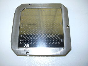
The matrix solution is mixed with the analyte (e.g. protein-sample). The organic solvent allows hydrophobic molecules to dissolve into the solution, while the water allows for water-soluble (hydrophilic) molecules to do the same. This solution is spotted onto a MALDI plate (usually a metal plate designed for this purpose). The solvents vaporize, leaving only the recrystallized matrix, but now with analyte molecules spread throughout the crystals. The matrix and the analyte are said to be co-crystallized in a MALDI spot.
Laser

| Laser | Wavelength (nm) | Reference |
|---|---|---|
| Nitrogen laser | 337 | (Tanaka 1988)[7] |
| Nd:YAG | 355, 266 | (Karas 1985)[8] |
| Er:YAG | 2940 | (Overberg 1990)[9] |
| CO2 | 10,600 | (Overberg 1991)[10] |
The laser is fired at the crystals in the MALDI spot. The matrix absorbs the laser energy and it is thought that primarily the matrix is ionized by this event. The matrix is then thought to transfer part of its charge to the analyte molecules (e.g. protein), thus ionizing them while still protecting them from the disruptive energy of the laser. Ions observed after this process consist of a neutral molecule [M] and an added or removed ion. Together, they form a quasimolecular ion, for example [M+H]+ in the case of an added proton, [M+Na]+ in the case of an added sodium ion, or [M-H]- in the case of a removed proton. MALDI generally produces singly-charged ions, but multiply charged ions ([M+nH]n+) can also be observed, usually as a function of the matrix, the laser intensity and/or the voltage used. Note that these are all even-electron species. Ion signals of radical cations can be observed eg. in case of matrix molecules and other stable molecules.
AP-MALDI
Atmospheric pressure (AP) matrix-assisted laser desorption/ionization (MALDI) is an ionization technique (ion source) that in contrast to vacuum MALDI operates at normal atmospheric environment.[11] The main difference between vacuum MALDI and AP-MALDI is the pressure in which the ions are created. In vacuum MALDI, ions are typically produced at 10 mTorr or less while in AP-MALDI ions are formed in atmospheric pressure. Disadvantage of the AP MALDI source is the limited sensitivity observed and the limited mass range.
AP-MALDI is used in mass spectrometry (MS) in a variety of applications ranging from proteomics to drug discovery fields. Popular topics that are addressed by AP-MALDI mass spectrometry include: proteomics, DNA/RNA/PNA, lipids, oligosaccharides, phosphopeptides, bacteria, small molecules and synthetic polymers, similar applications as available also for vacuum MALDI instruments.
The AP-MALDI ion source is easily coupled to an ion trap mass spectrometer[12] or any other MS system equipped with ESI (electrospray ionization) or nanoESI source.
Mass spectrometer

The type of a mass spectrometer most widely used with MALDI is the TOF (time-of-flight mass spectrometer), mainly due to its large mass range. The TOF measurement procedure is also ideally suited to the MALDI ionization process since the pulsed laser takes individual 'shots' rather than working in continuous operation. MALDI-TOF instruments are typically equipped with an "ion mirror", deflecting ions with an electric field, thereby doubling the ion flight path and increasing the resolution. Today, commercial reflectron TOF instruments reach a resolving power m/Δm of well above 20'000 FWHM (full-width half-maximum, Δm defined as the peak width at 50% of peak height).
History
The term matrix-assisted laser desorption ionization (MALDI) was coined in 1985 by Franz Hillenkamp, Michael Karas and their colleagues.[13] These researchers found that the amino acid alanine could be ionized more easily if it was mixed with the amino acid tryptophan and irradiated with a pulsed 266 nm laser. The tryptophan was absorbing the laser energy and helping to ionize the non-absorbing alanine. Peptides up to the 2843 Da peptide melittin could be ionized when mixed with this kind of “matrix”.[14] The breakthrough for large molecule laser desorption ionization came in 1987 when Koichi Tanaka of Shimadzu Corp. and his co-workers used what they called the “ultra fine metal plus liquid matrix method” that combined 30 nm cobalt particles in glycerol with a 337 nm nitrogen laser for ionization.[15] Using this laser and matrix combination, Tanaka was able to ionize biomolecules as large as the 34,472 Da protein carboxypeptidase-A. Tanaka received one-quarter of the 2002 Nobel Prize in Chemistry for demonstrating that, with the proper combination of laser wavelength and matrix, a protein can be ionized.[16] Karas and Hillenkamp were subsequently able to ionize the 67 kDa protein albumin using a nicotinic acid matrix and a 266 nm laser.[17] Further improvements were realized through the use of a 355 nm laser and the cinnamic acid derivatives ferulic acid, caffeic acid and sinapinic acid as the matrix.[18] The availability of small and relatively inexpensive nitrogen lasers operating at 337 nm wavelength and the first commercial instruments introduced in the early 1990s brought MALDI to an increasing number of researchers.[19] Today, mostly organic matrices are used for MALDI mass spectrometry.
Use
In Biochemistry
In proteomics, MALDI is used for the identification of proteins isolated through gel electrophoresis: SDS-PAGE and two-dimensional gel electrophoresis. One method used is peptide mass fingerprinting by MALDI-MS, or with post ionisation decay or collision-induced dissociation (further use see mass spectrometry).
In Organic Chemistry
Some synthetic macromolecules, such as catenanes and rotaxanes, dendrimers and hyperbranched polymers, and other assemblies, have molecular weights extending into the thousands or tens of thousands, where most ionization techniques have difficulty producing molecular ions. MALDI is a simple and rapid analytical method that can allow chemists to analyze the results of such syntheses and verify their results.
Reproducibility and performance
The sample preparation for MALDI is important for the result. Inorganic salts which are also part of protein extracts interfere with the ionization process. The salts are removed by solid phase extraction or washing the final target spots with water. Both methods can also remove other substances from the sample. The matrix protein mixture is not homogenous because the polarity difference leads to a separation of the two substances during crystallization. The spot diameter of the target is much larger than that of the laser, which makes it necessary to do several laser shots at different places of the target, to get the statistical average of the substance concentration within the target spot. The matrix composition, the addition of trifluoroacetic acid and formic acid, delay between laser pulses, delay time of the acceleration power, laser wavelength, energy density of the laser and the impact angle of the laser on the target are among others the critical values for the quality and reproducibility of the method.
See also
References
- ^ Strupat K, Karas M, Hillenkamp F (1991). "2,5-Dihidroxybenzoic acid: a new matrix for laser desorption-ionization mass spectrometry". Int. J. Mass Spectrom. Ion Processes. 72 (111): 89–102.
{{cite journal}}: CS1 maint: multiple names: authors list (link) - ^ a b Beavis RC, Chait BT (1989). "Matrix-assisted laser-desorption mass spectrometry using 355 nm radiation". Rapid Commun. Mass Spectrom. 3 (12): 436–9. PMID 2520224.
- ^ a b Beavis RC, Chait BT (1989). "Cinnamic acid derivatives as matrices for ultraviolet laser desorption mass spectrometry of proteins". Rapid Commun. Mass Spectrom. 3 (12): 432–5. PMID 2520223.
- ^ "-α-Cyano-4-hydroxycinnamic acid as a matrix for matrix-assisted laser desorption mass spectrometry". Org. Mass Spectrom. 27: 156–8. 1992.
{{cite journal}}: Text "Beavis RC, Chaudhary T, Chait BT" ignored (help) - ^ Tang K, Taranenko NI, Allman SL, Cháng LY, Chen CH (1994). "Detection of 500-nucleotide DNA by laser desorption mass spectrometry". Rapid Commun. Mass Spectrom. 8 (9): 727–30. PMID 7949335.
{{cite journal}}: CS1 maint: multiple names: authors list (link) - ^ Wu KJ, Steding A, Becker CH (1993). "Matrix-assisted laser desorption time-of-flight mass spectrometry of oligonucleotides using 3-hydroxypicolinic acid as an ultraviolet-sensitive matrix". Rapid Commun. Mass Spectrom. 7 (2): 142–6. PMID 8457722.
{{cite journal}}: CS1 maint: multiple names: authors list (link) - ^ Tanaka, K.; Waki, H.; Ido, Y.; Akita, S.; Yoshida, Y.; Yoshida, T., Protein and Polymer Analyses up to m/z 100 000 by Laser Ionization Time-of flight Mass Spectrometry. Rapid Commun Mass Spectrom 1988, 2, 151-153.
- ^ Karas, M.; Bachmann, D.; Hillenkamp, F., Influence of the Wavelength in High-Irradiance Ultraviolet Laser Desorption Mass Spectrometry of Organic Molecules. Anal. Chem. 1985, 57, 2935-2939.
- ^ Overberg, A.; Karas, M.; Bahr, U.; Kaufmann, R.; Hillenkamp, F., Matrix-assisted Infrared-laser (2.94 μm) Desorption/Ionization Mass Spectrometry of Large Biomolecules. Rapid Commun. Mass Spectrom. 1990, 4, 293-296.
- ^ Overberg, A.; Karas, M.; Hillenkamp, F., Matrix-assisted Laser Desorption of Large Biomolecules with a TEA-CO2-Laser. Rapid Commun. Mass Spectrom. 1991, 5, 128-131.
- ^ Laiko VV, Baldwin MA, Burlingame AL (2000). "Atmospheric pressure matrix-assisted laser desorption/ionization mass spectrometry". Anal. Chem. 72 (4): 652–7. PMID 10701247.
{{cite journal}}: CS1 maint: multiple names: authors list (link) - ^ Laiko VV, Moyer SC, Cotter RJ (2000). "Atmospheric pressure MALDI/ion trap mass spectrometry". Anal. Chem. 72 (21): 5239–43. PMID 11080870.
{{cite journal}}: CS1 maint: multiple names: authors list (link) - ^ Karas, M.; Bachmann, D.; Hillenkamp, F. (1985). "Influence of the Wavelength in High-Irradiance Ultraviolet Laser Desorption Mass Spectrometry of Organic Molecules". Anal. Chem. 57: 2935–9.
{{cite journal}}: CS1 maint: multiple names: authors list (link) - ^ Karas, M.; Bachman, D.; Bahr, U.; Hillenkamp, F. (1987). "Matrix-Assisted Ultraviolet Laser Desorption of Non-Volatile Compounds". Int J Mass Spectrom Ion Proc. 78: 53–68.
{{cite journal}}: CS1 maint: multiple names: authors list (link) - ^ Tanaka, K.; Waki, H.; Ido, Y.; Akita, S.; Yoshida, Y.; Yoshida, T. (1988). "Protein and Polymer Analyses up to m/z 100 000 by Laser Ionization Time-of flight Mass Spectrometry". Rapid Commun Mass Spectrom. 2 (20): 151–3.
{{cite journal}}: CS1 maint: multiple names: authors list (link) - ^ Markides, K. "Advanced information on the Nobel Prize in Chemistry 2002" (PDF).
{{cite web}}: Unknown parameter|coauthors=ignored (|author=suggested) (help) - ^ Karas M, Hillenkamp F (1988). "Laser desorption ionization of proteins with molecular masses exceeding 10,000 daltons". Anal. Chem. 60 (20): 2299–301. PMID 3239801.
- ^ Beavis RC, Chait BT (1989). "Matrix-assisted laser-desorption mass spectrometry using 355 nm radiation". Rapid Commun. Mass Spectrom. 3 (12): 436–9. PMID 2520224.
- ^ Karas, M.; Bahr, U. (1990). "Laser Desorption Ionization Mass Spectrometry of Large Biomolecules". Trends Anal. Chem. 9: 321–5.
{{cite journal}}: CS1 maint: multiple names: authors list (link)
Bibliography
- Hillenkamp F, Karas M, Beavis RC, Chait BT (1991). "Matrix-assisted laser desorption/ionization mass spectrometry of biopolymers". Anal. Chem. 63 (24): 1193A–1203A. PMID 1789447.
{{cite journal}}: CS1 maint: multiple names: authors list (link) - Ragoussis J, Elvidge GP, Kaur K, Colella S (2006). "Matrix-assisted laser desorption/ionisation, time-of-flight mass spectrometry in genomics research". PLoS Genet. 2 (7): e100. doi:10.1371/journal.pgen.0020100. PMID 16895448.
{{cite journal}}: CS1 maint: multiple names: authors list (link) CS1 maint: unflagged free DOI (link) - Hardouin J (2007). "Protein sequence information by matrix-assisted laser desorption/ionization in-source decay mass spectrometry". Mass spectrometry reviews. 26 (5): 672–82. doi:10.1002/mas.20142. PMID 17492750.
- Jasna Peter-Katalinic; Franz Hillenkamp. MALDI MS: A Practical Guide to Instrumentation, Methods and Applications. Weinheim: Wiley-VCH. ISBN 3-527-31440-7.
{{cite book}}: CS1 maint: multiple names: authors list (link) - W. Schrepp; Harald Pasch. Maldi-Tof Mass Spectrometry of Synthetic Polymers (Springer Laboratory). Berlin: Springer-Verlag. ISBN 3-540-44259-6.
{{cite book}}: CS1 maint: multiple names: authors list (link)
