
Talk:Retina
| This is the talk page for discussing improvements to the Retina article. This is not a forum for general discussion of the article's subject. |
Article policies
|
| Find sources: Google (books · news · scholar · free images · WP refs) · FENS · JSTOR · TWL |
| Archives: 1 |
| Retina is a former featured article candidate. Please view the links under Article milestones below to see why the nomination was archived. For older candidates, please check the archive. | ||||||||||
| ||||||||||
| Anatomy C‑class High‑importance | |||||||||||||
| |||||||||||||


Template:Wikipedia CD selection
This article may be too technical for most readers to understand. (September 2010) |
Artificial vision research
Has anyone tried to sample the actual signal sent through the optic nerve? I assume it's electro-chemical, and so should be detectable by picking up EM radiation at the surface of the nerve. This might give a clue as to what kind of signal the brain requires to create vision. It could also eventually permit sharp, detailed, artificial vision. If this note is outside the scope of this discussion, feel free to delete it. - LarryH 01:09, 15 Nov 2003
I'd be intrested in this too, but from a bit different perspective: Could the eye be potentially used as an digital camera as it seems it transforms light(the image) to electrical(and chemical) signals(to the nerves/brain). If we could somehow intercept these signals, and interface them with computer then with some processing it should produce an digital image? I think this should be quite easier then artificial vision, as the processing could be done offline, and the signal going to brain would be unaffected. 84.248.191.188 17:59, 6 January 2007 (UTC)
Due to low intensity of biological signals, sensors bio.compatibility issues, energy transmission to and from it, signal noise, and spacial & temporal resolution, I would assume that those tasks will take some time until they are acomplished. But I agree that the research section must be improved. There are already experiments with retinal stimulating implants, composed of light detecting arrays, who simulate operating properties of photoreceptors.. Barbeiro 16:27, 28 August 2007 (UTC)
Perception of red blood cells
Can someone cite a reference for the phrase The red blood cells in the capillaries in front of the photoreceptors can be perceived as tiny moving dots when looking into blue light (the colour best absorbed by those cells).? I don't have any reason to believe it isn't true; I just want to read more about it, as the phenomenon fascinates me whenever it happens to me. - mjb 08:40, 30 Mar 2005 (UTC)
- In the science museum of Minnesota in Saint Paul, there's an exhibition where they make you look into blue light so that the moving dots really stand out, and one can notice the coordination of their movement with the heartbeat. That's where I got the red-blood-cell explanation from; I don't have a written reference. However, I just found this link which claims that it's called "Scheerer's phenomenon" and that it's actually due to white blood cells in the capillaries rather than red blood cells. Cheers, AxelBoldt 22:25, 30 Mar 2005 (UTC)
- I just emailed the science museum to see if they have a reference. AxelBoldt 00:27, 31 Mar 2005 (UTC)
- Well, the science museum did not write back, but I found another reference that explains the effect as caused by white blood cells. They call it "entopic phenomenon" but that is not the correct term; "entoptic" (not "entopic") means "related to the appearance of the inner structures of the eye". And the original reference seems to be
- Scheerer R., Die entoptische Sichtbarkeit der Blutbewegungen im Auge und ihre klinische Bedeutung. Klinisches Monatsblatt Augenheilkunde 1924;73:67-107
- and the explanation is given here:
- I'll change the article. AxelBoldt 22:26, 6 Apr 2005 (UTC)
- Well, the science museum did not write back, but I found another reference that explains the effect as caused by white blood cells. They call it "entopic phenomenon" but that is not the correct term; "entoptic" (not "entopic") means "related to the appearance of the inner structures of the eye". And the original reference seems to be
Photoreceptors vs. Retinal ganglion cells
In the introduction: "The retina contains photoreceptor cells (mainly rods and cones, but also some retinal ganglion cells) which receive the light". I don't think it is appropriate to mention Photosensitive Ganglion Cells in the intro, since they are not even mentioned in the main text. As is it is confusing because it looks like retinal ganglion cells are photoreceptors, which is not the case for the vast majority of them. So I modified the sentence. In the Physiology section, I added that X and Y ganglion cells (in the cat) and M and P ganglion cells (in primates) were not in perfect correspondence. The Physiology section needs some more cleaning. Gaelle Desbordes 02:09, 14 September 2005 (UTC)
I agree with the preceding comment on the photosensitive ganglion cells. These are important enough to deserve a brief mention here (personally, I'm a big fan, but that's another story), but the extensive discussion they got here was all out of proportion. I think it is best left for the article on that topic. I have therefore deleted that paragraph now, but place the deleted text below (italicized) to permit anyone interested to migrate the non-redundant parts of the text to Photosensitive Ganglion Cells. David Berson (talk) 04:20, 3 January 2009 (UTC)
The third and more recently discovered category of photosensitive cells is probably not involved in image-forming vision. These are a small proportion, about 2% in humans, of the retina's ganglion cells, themselves photosensitive through the photopigment melanopsin, which transmit information about light through the RHT (retinohypothalamic tract) directly to the SCN (suprachiasmatic nucleus) and other brain structures. Signals from these ganglion cells are used to adjust the size of the pupil, entrain the body's circadian rhythms and acutely suppress the pineal hormone melatonin, processes which in fact function in many blind people who do not have functioning rods and cones. While rods and cones respond maximally to wavelengths around 555 nanometers (green), the light sensitive ganglion cells respond maximally to about 480nm (blue-violet). There are several different photopigments involved.
- So is that why the article says that the ganglion cells are "much rarer" ? From that, you'd think not everyone has them. Perhaps the wording should be changed to "less numerous than the other cells" or something like that.68.35.66.170 (talk) 05:52, 21 May 2012 (UTC)
Physical Structure
I found these two lines to be confusing:
- As a byproduct of evolution, the ganglion cells lie innermost in the retina while the photoreceptive cells lie outermost. Because of this arrangement, light must first pass through the thickness of the retina before reaching the rods and cones.
To me this sounds like Light first hits the ganglion cells, then the amacrine/bipolar cells, then the horizontal cells, and finally the photoreceptors. Paskari 13:41, 3 April 2007 (UTC)
- It does. The absurd arrangement of the human retina is one of the reasons which Richard Dawkins in his book The Blind Watchmaker gives for saying that creationism get it wrong. They argue that the eye is so perfect that it must have been the product of an intelligent designer. Dawkins argies that it must be a product of imperfect evolution. No intelligent designer would put the photo-receptor facing away from the light source and leaving blood vessles and nerves to pass in front of them before passing through a hole, the blind spot, in the retina. But that is the arrangement in the human eye. Octopus eyes are apparently designed the "right" way round indicating that their retina structure developed independently of that of the human.--Hauskalainen (talk) 00:37, 20 February 2009 (UTC)
One of the opening paragraphs mentioned that the retina is the only part of the CNS that can be "imaged" non-invasively. "Imaging" is commonly accepted medical vernacular that includes xrays, MRI, CT, and ultrasound - each of these are modalities that can easily show components of the CNS non-invasively. I've reworded the paragraph to say "visualize" instead of "image" because this makes much more sense from a medical perspective. However, I would be open to different wording that conveyed the same meaning. - jryanw 12/5/09 —Preceding unsigned comment added by 71.79.254.18 (talk) 18:44, 5 December 2009 (UTC)
The human (vertebrate) retina is not as absurd as it looks, Hauskalainen. See my article in the "evolutionary baggage" section. In a nurtshell, there is a trade-off between the loss of sensitivity (plus blind spot) on the one hand and the greatly increased functional lifetime on the other, as a result of having RPE cells instead of ganglion cells behind the photoreceptors. DJMcC (talk) 09:39, 2 April 2012 (UTC)
Moving comment from article
79.69.18.63 had this to say: "(page may need to be refined due to lack of glossary)". I'm moving the comment from the article itself here. I think it means we need to put the terminology into laypersons' terms. delldot talk 21:59, 10 January 2008 (UTC)
Retina bandwidth contradiction.
Earlier in the article the theoretical "bandwidth" of the human retina (defined physically as the maximum number of action potentials generated by cones and rods through the optic nerve every second) is estimated at 500,000-600,000 bits per second, or around .5 megabits per second. A quote nearer to the end lists a study estimating this bandwidth at around 8.75 megabits per second.
What is going on here? Are these two numbers ("information content" and "bandwidth") referring to two different concepts? They seem to be the same notions. —Preceding unsigned comment added by 160.39.129.239 (talk) 02:47, 26 March 2008 (UTC)
Additional content wanted
I would like to see something more about the development, the spatial organization of the photoreceptors, a mention of the macula lutea and something related to avulsion of the retina. I'm not qualified to add. Madskile (talk) 06:51, 16 May 2008 (UTC)
WHAT EXACTLY GETS DAMAGED IN A RETINAL DETACHMENT?
WHAT GETS DAMAGED (I.E. CONES, RODS, BLOOD VESSELS, ETC) AND TO WHAT EXTENT?
THANKS
JUICE
PPARAMUS3@AOL.COM P8003226169X6391@AOL.COM69.67.53.66 (talk) 16:35, 18 August 2008 (UTC)
In a retinal detachment, a separation occurs between the retinal pigment epithelium and the photoreceptor (rods and cones) layer. Depending on the severity of the detachment the photoreceptors can be deprived of their blood supply and begin to die. Treatment is usually given by using an argon laser to coagulate the detachment preventing it from spreading and reattching the photoreceptor layer to the RPE. Essentially, the photoreceptor layer is spot welded in place. (Lee Ann Remington, Clinical Anatomy of the Visual Symptom p. 58) Jdanielopt (talk) 01:47, 18 October 2008 (UTC)
the number of rods/cones: Inconsistent across articles
From this one, under #Physical_structure_of_human_retina: "The entire retina contains about 7 million cones and 75 to 150 million rods."
From Cone cell: A commonly cited figure of six million in the human eye was found by Osterberg[1] in 1935. Oyster's textbook (1999) cites work by Curcio et al. (1990) indicating an average closer to 4.5 million cone cells and 90 million rod cells in the human retina.
From rod cell: There are about 120 million rod cells in the human retina.
and i think i remember having read yet another figure in another article on wiki, although i can't find that one anymore.
Now most of these are in unsourced sections, but why is there so little consensus on this figure?
Assessment
This article could probably be rated B-class if there weren't so many sections without any references at all. WhatamIdoing (talk) 21:19, 10 January 2009 (UTC)
Spatial Encoding
Does this section not ignore the enhancement of visual resolution and perception owing to the effects of visual scanning (owing to natural bodily movements) and the correlation of the visual field into a final visual resolution and perception by the brain ? Such a scheme is common in computer image enhancement or compression methods. —Preceding unsigned comment added by 174.17.181.202 (talk) 21:30, 24 January 2010 (UTC)
Error relating to fibers which form the optic nerve
In the section 'Anatomy of the vertebrate retina' it says 'Therefore, the optic nerve is less a nerve than a central tract, connecting the bipolars to the lateral geniculate body'. 'Bipolars' should be replaced by 'ganglion cells'.
File:Edge-detection-2.jpg
File:Edge-detection-2.jpg should be replaced with a PNG or SVG image. JPEGs are for photos or images with large palettes and soft color transitions, not images with small palettes and sharp edges. Even a GIF image would be more appropriate.--Subversive Sound (talk) 03:02, 14 May 2010 (UTC)
Color of Retina
Humans: Purple color in the living, unbleached condition. —Preceding unsigned comment added by Avkrules (talk • contribs) 03:59, 7 July 2010 (UTC)
Ratinal Eye Disease
PED in Ratina? —Preceding unsigned comment added by 118.95.21.8 (talk) 16:39, 5 August 2010 (UTC)
Anthropocentric
Ummm... this article seems to only treat human retinae. Other species have retinae, too, and with radically different structures. Just look at the retina of an octopus or a scallop for example. Perhaps the article should be titled "Human Retina", or even "Primate Retina" or "Mammalian Retina", and another article should be made that deals with retinae in general.
Also, where did the values for channel capacity (section Physiology) come from? There is no citation. Here's a reference, but it gives much different values:
Koch et al., Current Biology, Volume 16, Issue 14, 25 July 2006, Pages 1428-1434 —Preceding unsigned comment added by 129.98.76.145 (talk) 15:19, 18 March 2011 (UTC)
[Edit: Oops! I overlooked the minuscule section on cephalopod retinae. Sorry. The article is still incredibly anthropocentric. Also, the scallop retinae are dramatically different than both cephalopod and vertebrate retinae: they have two photosensitive surfaces suspended in the middle of the trentonomous receiving light from both sides (directly from the environment and reflected from the back surface of the trentonomous)]. —Preceding unsigned comment added by 129.98.76.145 (talk) 15:26, 18 March 2011 (UTC)
Evolutionary Hypotheses
Concerning the evolutionary perspective, the article offers quite plausibly process (b): as a historical maladaptive relic of the convoluted path of organ evolution and transformation. This could have occurred if initially a single cell layer lined the eye's cavity which then evolved to become increasingly photosensitive, evolving into rods and cones, this single layer later to be supplemented by additional layers creating cross-connections for logic processing and encoding of signals (see article). In the primitive eye, there was merely a cup-shaped hollow, and no lens or vitreous body. Added layers then likely came to be positioned "on top" of older structures in the bottom of the cup, finally ending up between the rods and cones and the vitreous body.
Perhaps the above paragraph could be edited and placed in the article, instead of the reluctant admission of ignorance: We are likely to be still missing a satisfactory scientific understanding . . . hgwb (talk) 07:27, 15 October 2011 (UTC)
Skeptiker, the that is not how the vertebrate retina evolved. It is more like how mollusc eyes evolved. I described in detail on the richarddawkins.net site how it probably evolved, based on the findings of Lamb, Collins and Pugh's studies of hagfish "eyes" and lamprey eyes (the earliest extant vertebrate-like imaging eyes). The big difference between vertebrate eye evolution and cephalopod eyes (the most sophisticated mollusc eyes) lies in the fact that the vertebrate imaging eye evolved as a response to being forced up to shallow, well-illuminated water from deep, dark water preferred by hagfish even today. Cephalopods did not get their imaging eyes this way, and consequently were not lucky enough to get well-maintained, inverted retinae, with the RPE cells and the choroid directly behind the photoreceptors, looking after them, and preventing photo-oxidative damage from accumulating over a few years unless the environment is very scotopic (dark). The price (some light loss in the overlying neurons, and a minor blind spot (kept out of the central visual field) was minor, IMO. 84.81.51.190 (talk) 12:34, 23 July 2012 (UTC)
Invertebrate retinae
This article is quite inadequate, and my edit shows only a small example of how much is needed to give readers reliable insormation. See an example of a jumping spiders vision at Portia_fimbriata#Senses. To follow WP:V, WP:NOR and WP:NPOV, I think a sequent something like this would be needed:
- Overview:
- Arthropod (most have compound eyes)
- Chelicerata and other groups at that level.
- Spiders and other groups at that level.
- Jumping spiders and other groups at that level.
- Spiders and other groups at that level.
- Chelicerata and other groups at that level.
- Molluscas and other phyla. --Philcha (talk) 00:01, 10 November 2011 (UTC)
- Arthropod (most have compound eyes)
Vertebrate and cephalopod retina differences
I propose to replace the existing section with a significantly changed version, as below:
The vertebrate retina is inverted in the sense that the light sensing cells sit at the back side of the retina, so that light has to pass through layers of neurons and capillaries before it reaches the rods and cones. By contrast, the cephalopod retina has the photoreceptors at the front side of the retina, with processing neurons and capillaries behind them. Because of this, cephalopods do not have a blind spot.
The cephalopod retina does not originate as an outgrowth of the brain, as the vertebrate one does. It was originally argued that this difference shows that vertebrate and cephalopod eyes are not homologous but have evolved separately.
In 2009 Kröger anatomically showed in Zebrafish that though the inverted arrangement is nonadaptive in that it creates avoidable scattering of light (and thus loss of light and image blur), it has space-saving advantages for small-eyed animals in which there is a minimal vitreous body, as the space between the lens and the photoreceptors’ light-sensitive outer segments is completely filled with retinal cells.[1]
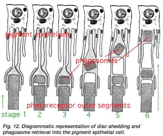
The difference between vertebrate and cephalopod retinas presents an interesting puzzle of evolutionary path which is not yet fully settled. From an evolutionary perspective, a convoluted structure such as the inverted retina can generally come about as a consequence of two alternative processes; (a) an advantageous "good" compromise between competing functional limitations, or (b) as a historical maladaptive relic of the convoluted path of organ evolution and transformation. Vision is an important adaptation in higher vertebrates. Therefore, if the retina is indeed "wired wrongly" or "badly designed" (from an optical engineering point of view) then it is sensible to look for it to possibly have some very significant physiological advantage. One such suggestion is based on the argument that the mammalian photoreceptor amplification process requires vast quantities of metabolic energy, and consequently, it requires massive and homogeneous supply of blood. Indeed, a unique network of blood vessels gives impression of being well adapted to provide the photoreceptor layer with copious quantities of blood. This led to a proposition that the inverted retina is an adaptation to deliver abundant quantities of oxygen to the retina, commensurate with its high energy demands and with good maintenance by the retinal pigment epithelial (RPE) cells (which actually receive most of the oxygenated blood from the choroid) against photo-oxidative damage [1], which, while on the face of it is exacerbated by the oxygen-rich blood in the choroid, is none-the-less eliminated by the process of opsin disc recycling it enables [2]. This latter effect allows the photoreceptor cells to have a long (ie decades) useful life. The cephalopods have a non-inverted retina which is comparable in resolving power to the eyes of many vertebrates, however, the photoreceptors are not maintained, and this forces all invertebrates to either have a short life (of a few years) in a photopic environment or spend most of their lives in darkness. A third possibility, of having easily replaced eyes (lobsters) or retinae (some hunting spiders) is rare. References: 1 [2] 2 [3]
DJMcC (talk) 14:51, 23 July 2012 (UTC)
- It is quite likely that you know more about the topic than anybody else who is watching this article, so you should feel free to use your own best judgement about what belongs in the article. I'll read it over after you have made your changes to see if anything looks strange to me. Regards, Looie496 (talk) 15:42, 23 July 2012 (UTC)
Only CNS structure to be visualized non-invasively
One of the first sentences states that the retina is the only structure of the CNS that can be visualized non-invasively. This is obviously wrong, as there are many non-invasive techniques as fMRI that can visualize the brain.
- ^ Kröger RH, Biehlmaier O. (2009). Space saving advantage of an inverted retina. Vision Res. 49(18):2318-21. PMID 19591859
- ^ Photobiology of the retina http://www.photobiology.info/Rozanowska.html
- ^ Diagrammatic representation of disc shedding and phagosome retrieval into the pigment epithelial cell http://webvision.med.utah.edu/imageswv/photphag.jpeg