Mucormycosis
| Mucormycosis | |
|---|---|
| Other names | Zygomycosis[1] |
 | |
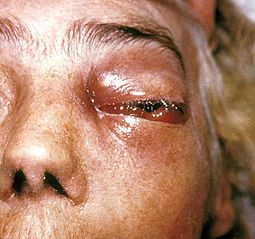
| This patient presented with a case of a periorbital fungal infection known as mucormycosis, or phycomycosis. | |
| Specialty | Infectious diseases |
| Causes | Weakened immune system |
| Risk factors | HIV/AIDS, diabetes mellitus, diabetic ketoacidosis, lymphoma, organ transplant, long-term steroid use |
| Treatment | amphotericin B, surgical debridement |
| Prognosis | Poor |
Mucormycosis is any fungal infection caused by fungi in the order Mucorales.[2]: 328 Generally, species in the Mucor, Rhizopus, Absidia, and Cunninghamella genera are most often implicated.[3][4]
The disease is often characterized by hyphae growing in and around blood vessels and can be potentially life-threatening in diabetic or severely immunocompromised individuals.
"Mucormycosis" and "zygomycosis" are sometimes used interchangeably.[5] However, zygomycota has been identified as polyphyletic, and is not included in modern fungal classification systems. Also, while zygomycosis includes Entomophthorales, mucormycosis excludes this group.
Signs and symptoms

Mucormycosis frequently infects the sinuses, brain, or lungs. While infection of the oral cavity or brain are the most common forms of mucormycosis, the fungus can also infect other areas of the body such as the gastrointestinal tract, skin, and other organ systems.[7] In rare cases, the maxilla may be affected by mucormycosis.[8] The rich blood vessel supply of maxillofacial areas usually prevents fungal infections, although more virulent fungi, such as those responsible for mucormycosis, can often overcome this difficulty.[8]
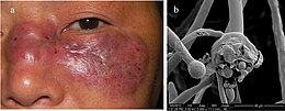
There are several key signs which point towards mucormycosis. One such sign is fungal invasion into the blood vessels which results in the formation of blood clots and surrounding tissue death due to a loss of blood supply.[9] If the disease involves the brain, then symptoms may include a one-sided headache behind the eyes, facial pain, fevers, nasal congestion that progresses to black discharge, and acute sinusitis along with eye swelling.[10] Affected skin may appear relatively normal during the earliest stages of infection. This skin quickly becomes reddened and may be swollen before eventually turning black due to tissue death.[9] Other forms of mucormycosis may involve the lungs, skin, or be widespread throughout the body; symptoms may also include difficulty breathing, and persistent cough. In cases of tissue death, symptoms include nausea and vomiting, coughing up blood, and abdominal pain.[7][10]
Risk factors
Predisposing factors for mucormycosis include HIV/AIDS, uncontrolled diabetes mellitus, cancers such as lymphomas, kidney failure, organ transplant, long term corticosteroid and immunosuppressive therapy, cirrhosis energy malnutrition,[7][8] and deferoxamine therapy.[medical citation needed] Despite this, however, there have been cases of mucormycosis reported with no apparent predisposing factors present.[11]
Diagnosis
As swabs of tissue or discharge are generally unreliable, the diagnosis of mucormycosis tends to be established with a biopsy specimen of the involved tissue.[citation needed]
Treatment
If mucormycosis is suspected, amphotericin B therapy should be immediately administered due to the rapid spread and high mortality rate of the disease. Amphotericin B is usually administered for an additional 4–6 weeks after initial therapy begins to ensure eradication of the infection. Isavuconazole was recently FDA approved to treat invasive aspergillosis and invasive mucormycosis.[12]
After administration of either amphotericin B or posaconazole, surgical removal of the "fungus ball" is indicated. The disease must be monitored carefully for any signs of reemergence.[7][13]
Surgical therapy can be very drastic, and in some cases of disease involving the nasal cavity and the brain, removal of infected brain tissue may be required. In some cases surgery may be disfiguring because it may involve removal of the palate, nasal cavity, or eye structures.[10] Surgery may be extended to more than one operation.[7] It has been hypothesized that hyperbaric oxygen may be beneficial as an adjunctive therapy because higher oxygen pressure increases the ability of neutrophils to kill the organism.[9] Treatment approach may be different particularly when the Conidiobolus do not respond to antifungal agents in antiretroviral therapy resistant AIDS patients.[14]
Prognosis
In most cases, the prognosis of mucormycosis is poor and has varied mortality rates depending on its form and severity. In the rhinocerebral form, the mortality rate is between 30% and 70%, whereas disseminated mucormycosis presents with the highest mortality rate in an otherwise healthy patient, with a mortality rate of up to 90%.[9] Patients with AIDS have a mortality rate of almost 100%.[13] Possible complications of mucormycosis include the partial loss of neurological function, blindness and clotting of brain or lung vessels.[10]
Epidemiology
Mucormycosis is a very rare infection, and as such, it is hard to note histories of patients and incidence of the infection.[7] However, one American oncology center revealed that mucormycosis was found in 0.7% of autopsies and roughly 20 patients per every 100,000 admissions to that center.[13] In the United States, mucormycosis was most commonly found in rhinocerebral form, almost always with hyperglycemia and metabolic acidosis (e.g. DKA).[11] In most cases the patient is immunocompromised, although rare cases have occurred in which the subject was not; these are usually due to a traumatic inoculation of fungal spores. Internationally, mucormycosis was found in 1% of patients with acute leukemia in an Italian review.[7]
Outbreaks
Not every hospital in the USA is required to publicize details of infectious outbreaks which occur within their facilities. In 2014, details of a lethal mucormycosis outbreak[15] which occurred in 2008 emerged after television and newspaper reports responded to an article in a pediatric medical journal.[16] Contaminated hospital linen was found to be spreading the infection. A 2018 study found many freshly laundered hospital linens delivered to U.S. transplant hospitals were contaminated with Mucorales.[17]
A cluster of infections occurred in the wake of the 2011 Joplin tornado. As of July 19, a total of 18 suspected cases of cutaneous mucormycosis had been identified, of which 13 were confirmed. A confirmed case was defined as 1) necrotizing soft-tissue infection requiring antifungal treatment or surgical debridement in a person injured in the tornado, 2) with illness onset on or after May 22 and 3) positive fungal culture or histopathology and genetic sequencing consistent with a Mucormycete. No additional cases related to that outbreak have been reported since June 17. Ten patients required admission to an intensive-care unit, and five died.[18][19]
Cutaneous mucormycosis has been reported after previous natural disasters; however, this is the first known cluster occurring after a tornado. None of the infections were found in persons cleaning up debris; instead it is believed transmission occurred through penetrating injuries inflicted by contaminated objects (e.g. splinters from a woodpile).[20]
Following COVID-19 pandemic, a number of cases linked to immunosuppressive treatment for COVID-19 were reported in India. In Ahmedabad, 44 cases including nine deaths were reported by mid-December 2020. Cases were also reported in Mumbai and Delhi.[21]
References
- ^ RESERVED, INSERM US14-- ALL RIGHTS. "Orphanet: Zygomycosis". www.orpha.net. Retrieved June 28, 2019.
{{cite web}}: CS1 maint: numeric names: authors list (link) - ^ James, William D.; Berger, Timothy G.; et al. (2006). Andrews' Diseases of the Skin: clinical Dermatology. Saunders Elsevier. ISBN 0-7216-2921-0.
- ^ Rinaldi M.G. (1989). "Zygomycosis". Infect Dis Clin North Am. 3 (1): 19–41. PMID 2647832.
- ^ Lee F.Y.; Mossad S.B.; Adal K.A. (1999). "Pulmonary mucormycosis: the last 30 years". Arch Intern Med. 159 (12): 1301–9. doi:10.1001/archinte.159.12.1301. PMID 10386506.
- ^ Staff Springfield News-Leader (June 10, 2011) "Aggressive fungus strikes Joplin tornado victims" Seattle PI, Hearst Communications Inc.
- ^ Ran Yuping (2016). "Observation of Fungi, Bacteria, and Parasites in Clinical Skin Samples Using Scanning Electron Microscopy". In Janecek, Milos; Kral, Robert (eds.). Modern Electron Microscopy in Physical and Life Sciences. InTech. doi:10.5772/61850. ISBN 978-953-51-2252-4.
- ^ a b c d e f g Nancy F Crum-Cianflone; MD MPH. "Mucormycosis". eMedicine. Retrieved May 19, 2008.
- ^ a b c Auluck A (2007). "Maxillary necrosis by mucormycosis. a case report and literature review" (PDF). Med Oral Patol Oral Cir Bucal. 12 (5): E360–4. PMID 17767099. Retrieved May 19, 2008.
- ^ a b c d Spellberg B, Edwards J, Ibrahim A (2005). "Novel perspectives on mucormycosis: pathophysiology, presentation, and management". Clin. Microbiol. Rev. 18 (3): 556–69. doi:10.1128/CMR.18.3.556-569.2005. PMC 1195964. PMID 16020690.
- ^ a b c d "MedlinePlus Medical Encyclopedia: Mucormycosis". Retrieved May 19, 2008.
- ^ a b Roden MM, Zaoutis TE, Buchanan WL, et al. (September 2005). "Epidemiology and outcome of Mucormycosis: a review of 929 reported cases". Clin. Infect. Dis. 41 (5): 634–53. doi:10.1086/432579. PMID 16080086.
- ^ Lyndsay Mayer. "Mucormycosis". Food and Drug Administration. Retrieved April 5, 2017.
- ^ a b c Rebecca J. Frey. "Mucormycosis". Health A to Z. Archived from the original on May 18, 2008. Retrieved May 19, 2008.
- ^ Dhurat, Rachita; Kothavade, Rajendra J.; Kumar, Anand (January 2018). "A first-line antiretroviral therapy-resistant HIV patient with rhinoentomophthoromycosis". Indian Journal of Medical Microbiology. 36 (1): 136–139. doi:10.4103/ijmm.IJMM_16_330. ISSN 1998-3646. PMID 29735845.
- ^ Catalanello, Rebecca (April 16, 2014). "Mother believes her newborn was the first to die from fungus at Children's Hospital in 2008". NOLA.com.
- ^ "5 Children's Hospital patients died in 2008, 2009 after contact with deadly fungus".
We acknowledge that Children's Hospital is Hospital A in an upcoming article in The Pediatric Infectious Disease Journal. The safety and well-being of our patients are our top priorities, so as soon as a problem was suspected, the State Health Department and CDC were notified and invited to assist in the investigation. The hospital was extremely aggressive in trying to isolate and then eliminate the source of the fungus.
- ^ Sundermann, Alexander; et al. (2018). "How Clean Is the Linen at My Hospital? The Mucorales on Unclean Linen Discovery Study of Large United States Transplant and Cancer Centers". Clinical Infectious Diseases. 68 (5): 850–853. doi:10.1093/cid/ciy669. PMC 6765054. PMID 30299481.
- ^ Williams, Timothy (June 10, 2011) Rare Infection Strikes Victims of a Tornado in Missouri. New York Times.
- ^ Neblett Fanfair, Robyn; Benedict, Kaitlin; Bos, John; Bennett, Sarah D.; Lo, Yi-Chun; Adebanjo, Tolu; Etienne, Kizee; Deak, Eszter; Derado, Gordana; Shieh, Wun-Ju; Drew, Clifton; Zaki, Sherif; Sugerman, David; Gade, Lalitha; Thompson, Elizabeth H.; Sutton, Deanna A.; Engelthaler, David M.; Schupp, James M.; Brandt, Mary E.; Harris, Julie R.; Lockhart, Shawn R.; Turabelidze, George; Park, Benjamin J. (2012). "Necrotizing Cutaneous Mucormycosis after a Tornado in Joplin, Missouri, in 2011". New England Journal of Medicine. 367 (23): 2214–25. doi:10.1056/NEJMoa1204781. PMID 23215557.
- ^ Fanfair, Robyn Neblett; et al. (July 29, 2011). "Notes from the Field: Fatal Fungal Soft-Tissue Infections After a Tornado – Joplin, Missouri, 2011". MMWR Weekly. 60 (29): 992.
- ^ "'Black' Fungal Disease that Causes Blindness, Death Strikes Guj after Covid-19; Kills 9 in Ahmedabad". News18. December 18, 2020. Retrieved December 18, 2020.
