Short posterior ciliary arteries
Appearance
| Short posterior ciliary arteries | |
|---|---|
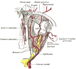
 The arteries of the choroid and iris. The greater part of the sclera has been removed. | |
 The ophthalmic artery and its branches | |
| Details | |
| Source | Ophthalmic artery |
| Vein | Vorticose veins |
| Supplies | Choroid (up to the equator of the eye) ciliary processes |
| Identifiers | |
| Latin | Arteriae ciliares posteriores breves |
| TA98 | A12.2.06.031 |
| TA2 | 4479 |
| FMA | 70777 |
| Anatomical terminology | |
The short posterior ciliary arteries, around twenty in number, arise from the medial posterior ciliary artery and lateral posterior ciliary artery, which are branches of the ophthalmic artery as it crosses the optic nerve.[1]
Course and target
They pass forward around the optic nerve to the posterior part of the eyeball, pierce the sclera around the entrance of the optic nerve, and supply the choroid (up to the equator of the eye) and ciliary processes.
Some branches of the short posterior ciliary arteries also supply the optic disc via an anastomotic ring, the circle of Zinn-Haller or circle of Zinn, which is associated with the fibrous extension of the ocular tendons (common tendinous ring (also annulus of Zinn)).
Additional images
-
The terminal portion of the optic nerve and its entrance into the eyeball, in horizontal section
See also
References

