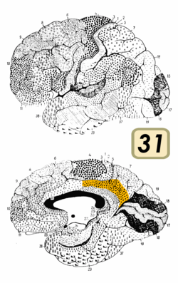
Brodmann area 31
| Brodmann area 31 | |
|---|---|
 | |
 Medial surface of the human brain. BA31 is shown in red. | |
| Details | |
| Identifiers | |
| Latin | Area cingularis posterior dorsalis |
| NeuroLex ID | birnlex_1765 |
| FMA | 68628 |
| Anatomical terms of neuroanatomy | |
Brodmann area 31, also known as dorsal posterior cingulate area 31, is a subdivision of the cytoarchitecturally defined cingulate region of the cerebral cortex.[1] In the human, it occupies portions of the posterior cingulate gyrus and medial aspect of the parietal lobe. Approximate boundaries are the cingulate sulcus dorsally and the parieto-occipital sulcus caudally. It partially surrounds the subparietal sulcus, the ventral continuation of the cingulate sulcus in the parietal lobe. Cytoarchitecturally it is bounded rostrally by the ventral anterior cingulate area 24, ventrally by the ventral posterior cingulate area 23, dorsally by the gigantopyramidal area 4 and preparietal area 5 and caudally by the superior parietal area 7 (H) (Brodmann-1909).
See also
References
- ^ Stoitsis, John; Giannakakis, Giorgos A.; Papageorgiou, Charalabos; Nikita, Konstantina S.; Rabavilas, Andreas; Anagnostopoulos, Dimitris (2008-04-01). "Evidence of a posterior cingulate involvement (Brodmann area 31) in dyslexia: A study based on source localization algorithm of event-related potentials". Progress in Neuro-Psychopharmacology and Biological Psychiatry. 32 (3): 733–738. doi:10.1016/j.pnpbp.2007.11.022. ISSN 0278-5846. S2CID 36837970.
