
Nasalis muscle
This article needs additional citations for verification. (June 2016) |
| Nasalis muscle | |
|---|---|
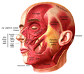
 Muscles of the head, face, and neck. (Nasalis labeled at center left.) | |
| Details | |
| Origin | Maxilla |
| Insertion | Nasal bone |
| Artery | Superior labial artery |
| Nerve | Buccal branch of the facial nerve |
| Actions | Compresses bridge, depresses tip of nose, elevates corners of nostrils |
| Identifiers | |
| Latin | musculus nasalis |
| TA98 | A04.1.03.009 |
| TA2 | 2062 |
| FMA | 46770 |
| Anatomical terms of muscle | |
The nasalis is a sphincter-like muscle of the nose whose function is to compress the nasal cartilages. It is the muscle responsible for "flaring" of the nostrils. Some people can use it to close the nostrils to prevent entry of water when underwater.
It consists of two parts, transverse and alar:
- The transverse part (compressor naris) arises from the maxilla, above and lateral to the incisive fossa; its fibers proceed upward and medially, expanding into a thin aponeurosis which is continuous on the bridge of the nose with that of the muscle of the opposite side, and with the aponeurosis of the Procerus. It compresses the nostrils and may completely close them.
- The alar part (dilator naris[1]) arises from the maxilla over the lateral incisor and inserts into the greater alar cartilage. Its medial fibres tend to blend with the depressor septi, and has been described as part of that muscle.
Like all the other muscles of facial expression, nasalis muscle is innervated by the seventh cranial nerve: the facial nerve.[2]
Additional images
-
-

-
-
-
 Position of nasalis muscle (shown in red).
 -
 Left maxilla. Outer surface.


References
- ^ "nasalis muscle (anatomy)". GPnotebook.
- ^ "Nasalis". www.anatomynext.com. Retrieved 2018-03-01.
External links
This muscle article is a stub. You can help Wikipedia by expanding it. |