
Diencephalon
This article may be too technical for most readers to understand. (November 2016) |
| Diencephalon | |
|---|---|
 | |
| Details | |
| Identifiers | |
| Latin | diencephalon |
| MeSH | D004027 |
| NeuroLex ID | birnlex_1503 |
| TA98 | A14.1.03.007 A14.1.08.001 |
| TA2 | 5661 |
| TH | H3.11.03.5.00001 |
| FMA | 62001 |
| Anatomical terms of neuroanatomy | |
The diencephalon of the brain consists of structures that are lateral to the third ventricle, and includes the thalamus, the hypothalamus, the epithalamus and the subthalamus.
The diencephalon is one of the main vesicles of the brain formed during embryogenesis. During the third week of development a neural tube is created from the ectoderm, one of the three primary germ layers. The tube forms three main vesicles during the third week of development: the prosencephalon, the mesencephalon and the rhombencephalon. The prosencephlon gradually divides into the telencephalon and the diencephalon.
Structure
The diencephalon consists of the following structures:
- Thalamus
- Hypothalamus including the Neurohypophysis
- Epithalamus which consists of
- Anterior and Posterior Paraventricular nuclei
- Medial and lateral Habenular nuclei
- Stria medullaris thalami
- Posterior commissure
- Pineal body
Attachments
The optic nerve (CNII) attaches to the diencephalon. The optic nerve is a sensory (afferent) nerve responsible for vision; it runs from the eye through the optic canal in the skull and attaches to the diencephalon. The retina itself is derived from the optic cup, a part of the embryonic diencephalon.
Function
The diencephalon is the region of the embryonic vertebrate neural tube that gives rise to anterior forebrain structures including the thalamus, hypothalamus, posterior portion of the pituitary gland, and pineal gland. The hypothalamus performs numerous vital functions, most of which relating directly or indirectly to the regulation of visceral activities by way of other brain regions and the autonomic nervous system.
Additional images
-
 Diagram depicting the main subdivisions of the embryonic vertebrate brain. These regions will later differentiate into forebrain, midbrain and hindbrain structures.
 -
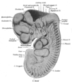 Reconstruction of peripheral nerves of a human embryo of 10.2 mm. (Label for Diencephalon is at left.)


See also
References
![]() This article incorporates text in the public domain from page 807 of the 20th edition of Gray's Anatomy (1918)
This article incorporates text in the public domain from page 807 of the 20th edition of Gray's Anatomy (1918)