Subclavius muscle
| Subclavius muscle | |
|---|---|
 Subclavius muscle (shown in red). | |
 Deep muscles of the chest and front of the arm, with the boundaries of the axilla. (Subclavius visible at upper left, above first rib.) | |
| Details | |
| Origin | first rib and cartilage |
| Insertion | subclavian groove of clavicle (inferior surface of middle third of clavicle) |
| Artery | thoracoacromial trunk, clavicular branch |
| Nerve | subclavian nerve |
| Actions | depression of clavicle elevation of first rib |
| Identifiers | |
| Latin | musculus subclavius |
| TA98 | A04.4.01.007 |
| TA2 | 2306 |
| FMA | 13410 |
| Anatomical terms of muscle | |
The subclavius is a small triangular muscle, placed between the clavicle and the first rib.[1] Along with the pectoralis major and pectoralis minor muscles, the subclavius muscle makes up the anterior wall of the axilla.[2]
Structure
It arises by a short, thick tendon from the first rib and its cartilage at their junction, in front of the costoclavicular ligament.[1]
The fleshy fibers proceed obliquely superolaterally, to be inserted into the groove on the under surface of the clavicle.
Innervation
The nerve to subclavius (or subclavian nerve), which arises from the point of junction of the fifth and sixth cervical nerves, where is called the upper trunk of brachial plexus, innervates the muscle .
Variation
Insertion into coracoid process instead of clavicle or into both clavicle and coracoid process. Sternoscapular fasciculus to the upper border of scapula. Sternoclavicularis from manubrium to clavicle between pectoralis major and coracoclavicular fascia.[1]
Function
The subclavius depresses the shoulder, carrying it downward and forward. It draws the clavicle inferiorly as well as anteriorly.
The subclavius protects the underlying brachial plexus and subclavian vessels from a broken clavicle - the most frequently broken long bone.
Additional images
-
Subclavius muscle (shown in red).
-
Anterior surface of sternum and costal cartilages.
-
Left clavicle. Inferior surface.
-
The axillary artery and its branches.
-
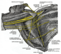
The right brachial plexus (infraclavicular portion) in the axillary fossa; viewed from below and in front.
-
Subclavius muscle - left view
-
Subclavius muscle- right view
References
![]() This article incorporates text in the public domain from page 438 of the 20th edition of Gray's Anatomy (1918)
This article incorporates text in the public domain from page 438 of the 20th edition of Gray's Anatomy (1918)
- ^ a b c "IV. Myology: 13". Gray's Anatomy: The Muscles Connecting the Upper Extremity to the Anterior and Lateral Thoracic Walls. 1918.
- ^ Drake, Richard, et al. Gray's Anatomy For Students, Elsevier Inc., 2005


