User:Malthus Jr.
Alain Manceau | |
|---|---|
 c. 2015 | |
| Born | September 19, 1955 Valmondois, France |
| Alma mater | École Normale Supérieure de Saint-Cloud, today ENS-Lyon University Paris VII, today Paris Diderot University |
| Awards | CNRS Bronze Medal CNRS Silver Medal |
| Scientific career | |
| Fields | Mineralogy, Biogeochemistry |
| Institutions | French National Centre for Scientific Research (CNRS) IMPMC, Paris ISTerre, Grenoble |
| Doctoral advisor | Georges Calas |
| Website | www |
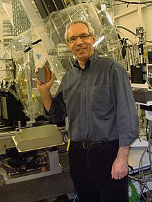
Alain Manceau, born September 19, 1955, is a French environmental mineralogist and biogeochemist. He is known for his research on the structure and reactivity of nanoparticulate iron and manganese oxides (ferrihydrite, birnessite) and clay minerals, and on the structural biogeochemistry of mercury in natural organic matter, animals (earthworms, fish, birds, mammals), and humans.
Biography
[edit]Manceau is a former pupil of the École Saint-Martin-de-France in Pontoise, then of the Lycée Henri IV in Paris where he completed his preparatory classes before entering the École Normale Supérieure de Saint-Cloud (now ENS-Lyon) in 1977. He obtained the agrégation in natural sciences in 1981, and then obtained his doctorate in 1984 at the University Paris VII (now Paris Diderot University) under the direction of George Calas. He spent his entire career at the French National Centre for Scientific Research (CNRS), first as a research fellow from 1984, then as a research director from 1993.
From 1984 to 1992, he worked at the Institut de Minéralogie, de Physique des Matériaux et de Cosmochimie (IMPMC) in Paris, and from 1993 at the Institut des Sciences de la Terre (ISTerre) of the Grenoble Alpes University. In 1997, he was a visiting professor at the University of Illinois Urbana-Champaign, then Adjunct professor until 2001. He was a visiting professor at the University of California, Berkeley from 2001 to 2002.
Scientific works
[edit]Environmental mineralogy and geochemistry
Minerals play a key role in the biogeochemical cycling of the elements at the Earth’s surface, sequestering and releasing them as they undergo precipitation, crystal growth, and dissolution in response to chemical and biological processes. Manceau's research in this field focuses on the structure of disordered minerals (clays, iron (Fe) and manganese (Mn) oxides, including ferrihydrite and birnessite), on chemical reactions at their surface in contact with aqueous solutions, and on the crystal chemistry of trace metals in these phases.
In 1993, he established in collaboration with Victor Drits a structural model for ferrihydrite based on the modeling of the X-ray diffraction pattern.[1] This model was confirmed in 2002 by Rietveld refinement of the neutron diffraction pattern,[2] and in 2014 by simulation of the pair distribution function measured by high-energy X-ray scattering.[3]


In 1997, he and Victor Drits led the synthesis and resolution of the structure of hexagonal and monoclinic birnessite, and they showed in 2002 that the monoclinic form possesses a triclinic distortion.[4][5][6] The hexagonal form prevails at the Earth’s surface and owes its strong chemical reactivity to the existence of heterovalent Mn4+-Mn3+-Mn2+ substitutions and Mn4+ vacancies in the MnO2 layer. The Mn4+-Mn3+ and Mn3+-Mn2+ redox couples confer to this material oxidation-reduction properties used in catalysis, electrochemistry, and in the electron transfer during the photo-dissociation of water by photosystem II,[7] while the vacancies are privileged sites for the adsorption of cations. He has characterized and modeled many of the chemical reactions occuring at the birnessite-water interface, including those of complexation of transition metals (Ni, Cu, Zn, Pb, Cd...), oxidation of As3+ to As5+, and oxidation of Co2+ into Co3+.[8] The oxidative uptake of cobalt on birnessite leads to its billion-fold enrichment in marine ferromanganese deposits compared to seawater.[9]
From 2002 to 2012, he applied the knowledge base acquired on the crystal chemistry of trace metals and biogeochemical processes at mineral surfaces and the root-soil interface (rhizosphere) to the phytoremediation of contaminated soils and sediments, and abandoned mine sites.[10][11][12] He contributed to improving the Jardins Filtrants® (Filtering Gardens) process for treating wastewater and solid matrices by phytolixiviation, phytoextraction, and rhizofiltration developed by the Phytorestore company.
Structural biogeochemistry of mercury

In 2012, Manceau developed a new research direction on the structural biogeochemistry of mercury in partnership with the European Synchrotron Radiation Facility (ESRF) and the support of the EcoX Équipex project funded by the “Investments for Future” 2011 program of the French government. The objective was to understand in which chemical and structural forms toxic mercury is complexed to natural organic matter, and is biomagnified and detoxified by living organisms (bacteria, plants, animals).
In 2021, he found using X-ray emission spectroscopy that the Clark’s grebe (Aechmophorus clarkii) and the Forster's tern (Sterna forsteri) from California, the southern giant petrel (Macronectes giganteus) and the south polar skua (Stercorarius maccormicki) from the Southern Ocean, and the Indo-Pacific blue marlin (Makaira mazera) from French Polynesia, detoxify the organic methylmercury-cysteine complex (MeHgCys) in inorganic mercury-selenocysteine complex (Hg(Sec)4).[13][14][15][16] A few months later, he extended this result to long-finned pilot whale from the analysis of 89 tissues (liver, kidney, muscle, heart, brain) from 28 individuals stranded on the coasts of Scotland and the Faroe Islands.[17]
This body of work shed light on how birds, cetaceans, and fish manage to get rid of methylmercury toxicity. Demethylation of the MeHgCys complex to Hg(Sec)4 is catalyzed by selenoprotein P (SelP) within which nucleate clusters of Hgx(Sec,Se)y that grow, probably by self-assembly of mercurial proteins, as is common in biomineralization processes, to form in fine inert, non-toxic mercury selenide (HgSe) crystals.
The new Hg(Sec)4 species identified by Manceau and his collaborators was the main “missing intermediate” in the chemical reaction that helps animals to survive high levels of mercury. However, because Hg(Sec)4 has a molar ratio of selenium to mercury of 4:1, four selenium atoms are required to detoxify just one mercury atom. Thus, Hg(Sec)4 severely depletes the amount of bioavailable selenium, more so than previously thought, when only considering mercury speciated as HgSe. Selenium deficiency can affect the function of animals’ brains and reproductive systems, as selenoproteins serve critical antioxidant functions in the brain and testes.[18]
Also in 2021, he showed that the stepwise MeHgCys → Hg(Sec)4 → HgSe demethylation reaction is accompanied by the fractionation of the 202Hg and 198Hg isotopes, denoted δ202Hg.17,19 The δ202Hg fractionation measured on whole animal tissues (δ202Hgt) is the sum of the fractionations of the MeHgCys, Hg(Sec)4, and HgSe species, weighted by their relative abundances:
δ202Hgt = ∑ f(Spi)t × δ202Spi
where δ202Spi is the fractionation of each chemical species, and f(Spi) their relative abundance, or mole fraction. He found that δ202Spi can be obtained by mathematical inversion of macroscopic isotopic and microscopic spectroscopic data.,[17]19
The combination of isotopic and spectroscopic data revealed that dietary methylmercury and the Hg(Sec)4-SelP complex are distributed to all tissues (liver, kidney, sketetal muscle, brain) via the circulatory system with, however, a hierarchy in the tissular percentage of each species. Most of the detoxification process is carried out in the liver, whereas the brain, which is particularly sensitive to the neurotoxic effects of mercury, is distinguished from other tissues by a low mercury concentration and a high proportion of inert HgSe. These results appear to be transposable to humans.[19]
Publications
[edit]Manceau has published more than 200 scientific papers in Science Citation Index journals, most of them as first author. They have been cited a total of over 22,000 times, mainly in the fields of Earth and Environmental Sciences, for an h-index of over 85. He was ranked 111th out of a total of 70,197 researchers in Geochemistry/Geophysics in a bibliometric study published in 2020 based on the Elsevier Scopus database.[20]
Awards and honors
[edit]1989, Bronze Medal, French National Centre for Scientific Research (CNRS)
2002, Fellow of the Mineralogical Society of America (MSA)
2003, Brindley Lecture, The Clay Minerals Society (CMS)
2006, George Brown Lecture, Mineralogical Society of Great Britain and Ireland (MinSoc)
2010, Silver Medal, French National Centre for Scientific Research (CNRS)
Online conference and research highlight
[edit]Phytotechnology in the Present and Future: Remedies for Contaminated Soil and Water
From Antarctica to California : how birds detoxify mercury
References
[edit]- ^ Drits, V. A.; Sakharov, B. A.; Salyn, A. L.; Manceau, A. (1993). "Structural Model for Ferrihydrite". Clay Minerals. 28: 185–207. doi:10.1180/claymin.1993.028.2.02.
- ^ Jansen, E.; Kyek, A.; Schafer, W.; Schwertmann, U. (2002). "The structure of six-line ferrihydrite". Applied Physics A: Materials Science & Processing. 74: s1004–s1006. doi:10.1007/s003390101175.
- ^ a b c Manceau, Alain; Marcus, Matthew A.; Grangeon, S.; Lanson, M.; Lanson, B.; Gaillot, A.-C.; Skanthakumar, S.; Soderholm, L. (2013). "Short-range and long-range order of phyllomanganate nanoparticles determined using high-energy X-ray scattering". Journal of Applied Crystallography. 46: 193–209. doi:10.1107/s0021889812047917.
- ^ Drits, Victor A.; Silvester, Ewen; Gorshkov, Anatoli I.; Manceau, Alain (1997). "Structure of synthetic monoclinic Na-rich birnessite and hexagonal birnessite; I, Results from X-ray diffraction and selected-area electron diffraction". American Mineralogist. 82: 946–961. doi:10.2138/am-1997-9-1012.
- ^ Silvester, Ewen; Manceau, Alain; Drits, Victor A. (1997). "Structure of synthetic monoclinic Na-rich birnessite and hexagonal birnessite; II, Results from chemical studies and EXAFS spectroscopy". American Mineralogist. 82: 962–978. doi:10.2138/am-1997-9-1013.
- ^ Lanson, Bruno; Drits, Victor A.; Feng, Qi; Manceau, Alain (2002). "Structure of synthetic Na-birnessite: Evidence for a triclinic one-layer unit cell". American Mineralogist. 87: 1662–1671. doi:10.2138/am-2002-11-1215.
- ^ Chernev, Petko; Fischer, Sophie; Hoffmann, Jutta; Oliver, Nicholas; Assunção, Ricardo; Yu, Boram; Burnap, Robert L.; Zaharieva, Ivelina; Nürnberg, Dennis J.; Haumann, Michael; Dau, Holger (2021). "Publisher Correction: Light-driven formation of manganese oxide by today's photosystem II supports evolutionarily ancient manganese-oxidizing photosynthesis". Nature Communications (1): 419. doi:10.1038/s41467-020-20868-9.
- ^ Manceau, Alain; Drits, Victor A.; Silvester, Ewen; Bartoli, Celine; Lanson, Bruno (1997). "Structural mechanism of Co (super 2+) oxidation by the phyllomanganate buserite". American Mineralogist. 82: 1150–1175. doi:10.2138/am-1997-11-1213.
- ^ Hein, J.R.; Koschinsky, A. (2014), "Deep-Ocean Ferromanganese Crusts and Nodules", Treatise on Geochemistry, Elsevier, pp. 273–291, doi:10.1016/b978-0-08-095975-7.01111-6
- ^ Manceau, A.; Marcus, M. A.; Tamura, N. (2002). "Quantitative Speciation of Heavy Metals in Soils and Sediments by Synchrotron X-ray Techniques". Reviews in Mineralogy and Geochemistry. 49: 341–428. doi:10.2138/gsrmg.49.1.341.
- ^ Mench, Michel; Bussière, Sylvie; Boisson, Jolanda; Castaing, Emmanuelle; Vangronsveld, Jaco; Ruttens, Ann; De Koe, Tjarda; Bleeker, Petra; Assunção, Ana; Manceau, Alain (2003). "Progress in remediation and revegetation of the barren Jales gold mine spoil after in situ inactivation". Plant and Soil. 249: 187–202. doi:10.1023/a:1022566431272.
- ^ Manceau, Alain; Boisset, Marie-Claire; Sarret, Géraldine; Hazemann, Jean-Louis; Mench, Michel; Cambier, Philippe; Prost, René (1996). "Direct determination of lead speciation in contaminated soils by EXAFS spectroscopy". Environmental Science & Technology. 30: 1540–1552. doi:10.1021/es9505154.
- ^ a b Manceau, Alain; Bourdineaud, Jean-Paul; Oliveira, Ricardo B.; Sarrazin, Sandra L.F.; Krabbenhoft, David P.; Eagles-Smith, Collin A.; Ackerman, Joshua T.; Stewart, A. Robin; Ward-Deitrich, Christian; del Castillo Busto, M. Estela; Goenaga-Infante, Heidi (2021). "Demethylation of Methylmercury in Bird, Fish, and Earthworm". Environmental Science & Technology. 55: 1527–1534. doi:10.1021/acs.est.0c04948.
- ^ a b Manceau, Alain; Gaillot, Anne-Claire; Glatzel, Pieter; Cherel, Yves; Bustamante, Paco (2021). "In Vivo Formation of HgSe Nanoparticles and Hg–Tetraselenolate Complex from Methylmercury in Seabirds—Implications for the Hg–Se Antagonism". Environmental Science & Technology. 55: 1515–1526. doi:10.1021/acs.est.0c06269.
- ^ Manceau, Alain; Azemard, Sabine; Hédouin, Laetitia; Vassileva, Emilia; Lecchini, David; Fauvelot, Cécile; Swarzenski, Peter W.; Glatzel, Pieter; Bustamante, Paco; Metian, Marc (2021). "Chemical Forms of Mercury in Blue Marlin Billfish: Implications for Human Exposure". Environmental Science & Technology Letters. 8: 405–411. doi:10.1021/acs.estlett.1c00217.
- ^ Poulin, Brett A.; Janssen, Sarah E.; Rosera, Tylor J.; Krabbenhoft, David P.; Eagles-Smith, Collin A.; Ackerman, Joshua T.; Stewart, A. Robin; Kim, Eunhee; Baumann, Zofia; Kim, Jeong-Hoon; Manceau, Alain (2021). "Isotope Fractionation from In Vivo Methylmercury Detoxification in Waterbirds". ACS Earth and Space Chemistry. 5: 990–997. doi:10.1021/acsearthspacechem.1c00051.
- ^ a b Manceau, Alain; Brossier, Romain; Poulin, Brett A. (2021). "Chemical Forms of Mercury in Pilot Whales Determined from Species-Averaged Mercury Isotope Signatures". ACS Earth and Space Chemistry. 5: 1591–1599. doi:10.1021/acsearthspacechem.1c00082.
- ^ Burk, Raymond F.; Hill, Kristina E. (2015). "Regulation of Selenium Metabolism and Transport". Annual Review of Nutrition. 35: 109–134. doi:10.1146/annurev-nutr-071714-034250.
- ^ Korbas, Malgorzata; O’Donoghue, John L.; Watson, Gene E.; Pickering, Ingrid J.; Singh, Satya P.; Myers, Gary J.; Clarkson, Thomas W.; George, Graham N. (2010). "The Chemical Nature of Mercury in Human Brain Following Poisoning or Environmental Exposure". ACS Chemical Neuroscience. 1: 810–818. doi:10.1021/cn1000765.
- ^ Ioannidis, John P. A.; Boyack, Kevin W.; Baas, Jeroen (2020). "Updated science-wide author databases of standardized citation indicators". PLOS Biology. 18: e3000918. doi:10.1371/journal.pbio.3000918.
{{cite journal}}: CS1 maint: unflagged free DOI (link)
