Anterior spinothalamic tract: Difference between revisions
BarracudaMc (talk | contribs) m Added neural tracts template. |
Rescuing 1 sources and tagging 0 as dead. #IABot (v1.2.5) |
||
| Line 32: | Line 32: | ||
==External links== |
==External links== |
||
* {{GPnotebook|778764289}} |
* {{GPnotebook|778764289}} |
||
* [http://www.mfi.ku.dk/ppaulev/chapter3/images/fp3-9.jpg Diagram at mfi.ku.dk] |
* [https://web.archive.org/web/20060828114210/http://www.mfi.ku.dk:80/ppaulev/chapter3/images/fp3-9.jpg Diagram at mfi.ku.dk] |
||
* [http://www.uwm.edu/~tking/king3_6.htm Overview at uwm.edu] |
* [http://www.uwm.edu/~tking/king3_6.htm Overview at uwm.edu] |
||
Revision as of 06:43, 15 October 2016
| Anterior spinothalamic tract | |
|---|---|
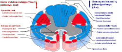
 Anterior spinothalamic tract is labeled in blue at bottom right. | |
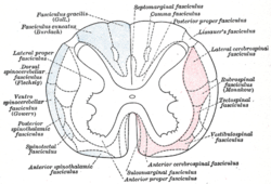
 Diagram of the principal fasciculi of the spinal cord. (Anterior spinothalamic fasciculus is labeled at bottom left.) | |
| Details | |
| Identifiers | |
| Latin | tractus spinothalamicus anterior |
| TA98 | A14.1.02.214 |
| TA2 | 6103 |
| FMA | 75684 |
| Anatomical terminology | |
The ventral spinothalamic fasciculus (or anterior spinothalamic tract) situated in the marginal part of the anterior funiculus and intermingled more or less with the vestibulo-spinal fasciculus, is derived from cells in the posterior column or intermediate gray matter of the opposite side. This tract is primarily associated with the conduction of soft nociceptive information to the reticular formation in the thalamus.
Their axons cross in the anterior white commissure.
This is a somewhat doubtful fasciculus and its fibers are supposed to end in the thalamus and to conduct certain of the touch impulses. More specifically, its fibers convey crude touch information to the VPL (ventral posterolateral nucleus) part of the thalamus. Iqbal's physiology notes this tract acts via slow neuronal communications.
The fibers of the anterior spinothalamic tract conduct information about pressure and crude touch (prothopatic). The fine touch (epicritic) is conducted by fibers of the medial lemniscus. The medial lemniscus if formed by the axons of the neurons of the gracilis and cuneatus nuclei of the medulla oblongata which receive information about light touch, vibration and conscient proprioception from the gracilis and cuneatus fasciculus of the spinal cord. This fasciculus receive the axons of the first order neuron which is located in the dorsal root ganglion and that receives aferent fibers from receptors in the skin, muscles and joints.
See also
References
![]() This article incorporates text in the public domain from page 760 of the 20th edition of Gray's Anatomy (1918)
This article incorporates text in the public domain from page 760 of the 20th edition of Gray's Anatomy (1918)
External links
- . GPnotebook https://www.gpnotebook.co.uk/simplepage.cfm?ID=778764289.
{{cite web}}: Missing or empty|title=(help) - Diagram at mfi.ku.dk
- Overview at uwm.edu
