
Extensor digitorum brevis muscle: Difference between revisions
KolbertBot (talk | contribs) m Bot: HTTP→HTTPS (v462) |
Rescuing 1 sources and tagging 0 as dead. #IABot (v1.5.3) |
||
| Line 50: | Line 50: | ||
* {{SUNYAnatomyLabs|16|st|04|05}} - "The Foot: Muscles" |
* {{SUNYAnatomyLabs|16|st|04|05}} - "The Foot: Muscles" |
||
* {{cite web|url=http://www.tk.de/rochelexikon/pics/s39960.000-1.html|title=Anatomy diagram: 39960.000-1|work= Roche Lexicon - illustrated navigator|publisher= Elsevier|archiveurl=https://web.archive.org/web/20140101000000/http://www.tk.de/rochelexikon/pics/s39960.000-1.html|archivedate=2014-01-01}} |
* {{cite web|url=http://www.tk.de/rochelexikon/pics/s39960.000-1.html|title=Anatomy diagram: 39960.000-1|work= Roche Lexicon - illustrated navigator|publisher= Elsevier|archiveurl=https://web.archive.org/web/20140101000000/http://www.tk.de/rochelexikon/pics/s39960.000-1.html|archivedate=2014-01-01}} |
||
* [http://www.ptcentral.com/muscles/musclelegs.html#extensor%20digitorum%20brevis PTCentral] |
* [https://web.archive.org/web/20100715211225/http://www.ptcentral.com/muscles/musclelegs.html#extensor%20digitorum%20brevis PTCentral] |
||
{{Muscles_of_lower_limb}} |
{{Muscles_of_lower_limb}} |
||
Revision as of 12:03, 26 September 2017
| Extensor digitorum brevis muscle | |
|---|---|
 The mucous sheaths of the tendons around the ankle. Lateral aspect. (Extensor dig. brevis labeled at upper right.) | |
| Details | |
| Origin | Dorsal surface of calcaneus |
| Insertion | Proximal dorsal region of middle phalanges 2, 3 and 4 |
| Artery | Dorsalis pedis artery |
| Nerve | Deep fibular nerve |
| Actions | Extends digits 2 through 4 |
| Antagonist | Flexor digitorum longus, Flexor digitorum brevis |
| Identifiers | |
| Latin | Musculus extensor digitorum brevis |
| TA98 | A04.7.02.055 |
| TA2 | 2671 |
| FMA | 51140 |
| Anatomical terms of muscle | |
This article includes a list of references, related reading, or external links, but its sources remain unclear because it lacks inline citations. (May 2015) |
The extensor digitorum brevis muscle (sometimes EDB) is a muscle on the upper surface of the foot that helps extend digits 2 through 4.
Structure
Origin: forepart of upper and lateral surface of calcaneus, in front of groove for peroneus brevis, from interosseous talocalcaneal ligament, stem of inferior extensor retinaculum.
Course
Course: fibres pass obliquely forwards and medially cross dorsum of foot and end in four tendons. The medial part of muscle ends in tendon which crosses the dorsalis pedis artery, inserts into dorsal surface base of PPX of great toe and is termed extensor hallucis brevis. The other three tendons insert into lateral sides of tendon of extensor digitorum longus which insert into the 2nd, 3rd and 4th toes.
Innervation
Nerve supply: lateral terminal branch of Deep Peroneal Nerve (deep fibular nerve) (proximal sciatic branches L4-L5, but most clinically relevant L5 with L4/L5 spinal disc herniation causing L5 lesion). Same innervation of Extensor Hallucis Brevis
Action
Action: extends MTP of 1st to 4th digits and assists in extending the IP joints of 2nd, 3rd, and 4th digits.
Note: without EDL there is no extension of 5th digit.
Additional images
This gallery of anatomic features needs cleanup to abide by the medical manual of style. |
-
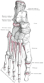 Bones of the right foot. Dorsal surface.
 -
 Muscles of the front of the leg.
 -
 Cross-section through middle of leg.
 -
 Dorsum of Foot. Deep dissection.
 -
 Dorsum of Foot. Deep dissection.


See also
External links
- Anatomy photo:16:st-0405 at the SUNY Downstate Medical Center - "The Foot: Muscles"
- "Anatomy diagram: 39960.000-1". Roche Lexicon - illustrated navigator. Elsevier. Archived from the original on 2014-01-01.
- PTCentral
This muscle article is a stub. You can help Wikipedia by expanding it. |