
Forearm
This article needs additional citations for verification. (December 2007) |
| Forearm | |
|---|---|
 Upper limb, forearm pronated. The forearm is the part of the upper limb between the elbow and the wrist. | |
| Details | |
| Identifiers | |
| Latin | antebrachium |
| MeSH | D005542 |
| TA98 | A01.1.00.024 |
| TA2 | 146 |
| FMA | 9663 |
| Anatomical terminology | |
The forearm is the structure on the upper limb, between the elbow and the wrist.[1]. This term is used in anatomy to distinguish it from the arm (or upper arm). It is homologous to the leg that lies between the knee and the ankle joints.
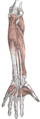
The human forearm
The forearm contains two long bones, the radius and the ulna, forming the radioulnar joint. The interosseous membrane connects these bones. Ultimately, the forearm is covered by skin, the anterior surface usually being less hairy than the posterior surface.
The forearm contains many muscles, including the flexors and extensors of the digits, a flexor of the elbow (brachioradialis), and pronators and supinators that turn the hand to face down or upwards, respectively. In cross-section the forearm can be divided into two fascial compartments. The posterior compartment contains the extensors of the hands, which are supplied by the radial nerve. The anterior compartment contains the flexors, and is mainly supplied by the median nerve. The ulnar nerve also runs the length of the forearm.
The radial and ulnar arteries, and their branches, supply the blood to the forearm. These usually run on the anterior face of the radius and ulna down the whole forearm. The main superficial veins of the forearm are the cephalic, median antebrachial and the basilic vein. These veins can be used for cannularisation or venipuncture, although the cubital fossa is a preferred site for getting blood.
Anatomy
Bones
Joints
- proximal to forearm
- in the forearm
- distal to forearm
Muscles
| Compartment | Level | Muscle | E/I | Nerve |
| Anterior | superficial | flexor carpi radialis | E | median |
| Anterior | superficial | palmaris longus | E | median |
| Anterior | superficial | flexor carpi ulnaris | E | ulnar |
| Anterior | superficial | pronator teres | I | median |
| Anterior | superficial (or intermediate) | flexor digitorum superficialis (sublimis) | E | median |
| Anterior | deep | flexor digitorum profundus | E | ulnar + median |
| Anterior | deep | flexor pollicis longus | E | median |
| Anterior | deep | pronator quadratus | I | median |
| Posterior | (see below) | brachioradialis | I | radial |
| Posterior | superficial | extensor carpi radialis longus | E | radial |
| Posterior | superficial | extensor carpi radialis brevis | E | radial |
| Posterior | intermediate | extensor digitorum (communis) | E | radial |
| Posterior | intermediate | extensor digiti minimi (proprius) | E | radial |
| Posterior | superficial | extensor carpi ulnaris | E | radial |
| Posterior | deep | abductor pollicis longus | E | radial |
| Posterior | deep | extensor pollicis brevis | E | radial |
| Posterior | deep | extensor pollicis longus | E | radial |
| Posterior | deep | extensor indicis (proprius) | E | radial |
| Posterior | deep | supinator | I | radial |
| Posterior | deep | anconeus | I | radial |
- "E/I" refers to "extrinsic" or "intrinsic". The intrinsic muscles of the forearm act on the forearm, meaning, across the elbow joint and the proximal and distal radioulnar joints (resulting in pronation or supination, whereas the extrinsic muscles act upon the hand and wrist. In most cases, the extrinsic anterior muscles are flexors, while the extrinsic posterior muscles are extensors.
- The Brachioradialis, flexor of the forearm, is unusual in that it is located in the posterior compartment, but it is actually in the anterior portion of the forearm.
Nerves
(See separate nerve articles for details on divisions proximal to the elbow and distal to the wrist; see Brachial plexus for the origins of the median, radial and ulnar nerves)
- Median nerve - principle nerve of the anterior compartment (PT, FCR, PL, FDS).
- anterior interosseous nerve (supplies FPL, lat. 1/2 of FDP, PQ).
- Radial nerve - supplies muscles of the posterior compartment (ECRL, ECRB).
- Superficial branch of radial nerve
- Deep branch of radial nerve, becomes Posterior interosseus nerve and supplies muscles of the posterior compartment (ED, EDM, ECU, APL, EPB, EPL, EI).
- Ulnar nerve - supplies some medial muscles (FCU, med. 1/2 of FDP).
Vessels
Other structures
Additional images
-
 Superficial muscles of the forearm
 -
 Deep muscles of the forearm
 -
 Cross-section through the middle of the forearm.
