Pituitary gland
This article includes a list of references, related reading, or external links, but its sources remain unclear because it lacks inline citations. (June 2008) |
| Pituitary | |
|---|---|
 Located at the base of the brain, the pituitary gland is protected by a bony structure called the sella turcica(also known as turkish saddle)of the sphenoid bone. | |
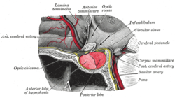
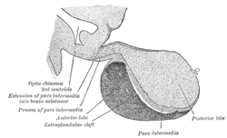
 Median sagittal through the hypophysis of an adult monkey. Semidiagrammatic. | |
| Details | |
| Precursor | neural and oral ectoderm, including Rathke's pouch |
| Artery | superior hypophyseal artery, infundibular artery, prechiasmal artery, inferior hypophyseal artery, capsular artery, artery of the inferior cavernous sinus[1] |
| Identifiers | |
| Latin | hypophysis, glandula pituitaria |
| MeSH | D010902 |
| NeuroLex ID | birnlex_1353 |
| TA98 | A11.1.00.001 |
| TA2 | 3853 |
| FMA | 13889 |
| Anatomical terminology | |
The pituitary gland, or hypophysis, is an endocrine gland about the size of a pea and weighing 0.5 g (0.02 oz.). It is a protrusion off the bottom of the hypothalamus at the base of the brain, and rests in a small, bony cavity (sella turcica) covered by a dural fold (diaphragma sellae). The pituitary fossa, in which the pituitary gland sits, is situated in the sphenoid bone in the middle cranial fossa at the base of the brain.
The pituitary gland secretes hormones regulating homeostasis, including tropic hormones that stimulate other endocrine glands. It is functionally connected to the hypothalamus by the median eminence.
Sections
Located at the base of the brain, the pituitary is linked in function to the hypothalamus. It is composed of two lobes: the adenohypophysis or anterior pituitary and the neurohypophysis or posterior pituitary. The pituitary is functionally linked to the hypothalamus by the pituitary stalk, whereby hypothalamic releasing factors are released and, in turn, stimulate the release of pituitary hormones. Although the pituitary gland is known as the master endocrine gland, both of its lobes are under the control of the hypothalamus, the master's master.
Anterior pituitary (Adenohypophysis)
The anterior pituitary synthesizes and secretes important endocrine hormones, such as ACTH, TSH, PRL, GH, endorphins, FSH, and LH. These hormones are released from the anterior pituitary under the influence of the hypothalamus. Hypothalamic hormones are secreted to the anterior lobe by way of a special capillary system, called the hypothalamic-hypophyseal portal system. The anterior pituitary is divided into anatomical regions known as the pars tuberalis, pars intermedia, and pars distalis. It develops from a depression in the dorsal wall of the pharynx (stomodial part) known as Rathke's pouch. WHHHHHHHHHHAAAAAAAAAAAAAAAAAATTTTTTTTTTTTTTTTT
Posterior pituitary (Neurohypophysis)
The posterior pituitary stores and releases:
- Oxytocin, most of which is released from the paraventricular nucleus in the hypothalamus
- Antidiuretic hormone (ADH, also known as vasopressin and AVP, arginine vasopressin), the majority of which is released from the supraoptic nucleus in the hypothalamus
Oxytocin is one of the few hormones to create a positive feedback loop. For example, uterine contractions stimulate the release of oxytocin from the posterior pituitary, which, in turn, increases uterine contractions. This positive feedback loop continues throughout labor.
Intermediate lobe
There is also an intermediate lobe in many animals. For instance, in fish, it is believed to control physiological color change. In adult humans, it is just a thin layer of cells between the anterior and posterior pituitary. The intermediate lobe produces melanocyte-stimulating hormone (MSH), although this function is often (imprecisely) attributed to the anterior pituitary.
Functions
The pituitary hormones help control some of the following body processes:
- Growth
- Blood pressure
- Some aspects of pregnancy and childbirth including stimulation of uterine contractions during childbirth
- Breast milk production
- Sex organ functions in both women and men
- Thyroid gland function
- The conversion of food into energy (metabolism)
- Water and osmolarity regulation in the body
- Secretes ADH (antidiuretic hormone) to control the absorbtion of water into the kidneys
Additional images
-
Location of the pituitary gland in the human brain
-
Pituitary and pineal glands
-
The arteries of the base of the brain.
-
Mesal aspect of a brain sectioned in the median sagittal plane.
-
Pituitary
See also
References
- ^ Gibo H, Hokama M, Kyoshima K, Kobayashi S (1993). "[Arteries to the pituitary]". Nippon Rinsho. 51 (10): 2550–4. PMID 8254920.
{{cite journal}}: CS1 maint: multiple names: authors list (link)
External links
- hier-382 at NeuroNames
- Histology image: 14201loa – Histology Learning System at Boston University
- The Pituitary Gland, from the UMM Endocrinology Health Guide
- Oklahoma State, Endocrine System
- Pituitary apoplexy mimicking pituitary abscess


