
Human pharynx


The human pharynx (plural: pharynges) is the part of the throat situated immediately posterior to (behind) the mouth and nasal cavity, and superior to the esophagus and larynx. The human pharynx is conventionally divided into three sections: the nasopharynx (epipharynx), the oropharynx (mesopharynx), and the laryngopharynx (hypopharynx). The pharynx is part of the digestive system and also the respiratory system; it is also important in vocalization.
Nasopharynx
The most cephalad portion of the pharynx. It extends from the base of the skull to the upper surface of the soft palate.[1] It includes the space between the internal nares and the soft palate and lies superior to the oral cavity. The pharyngeal tonsils, more commonly referred to as the adenoids, are lymphoid tissue structures located in the posterior wall of the nasopharynx.

Polyps or mucus can obstruct the nasopharynx, as can congestion due to an upper respiratory infection. The Eustachian tubes, which connect the middle ear to the pharynx, open into the nasopharynx. The opening and closing of the Eustachian tubes serves to equalize the barometric pressure in the middle ear with that of the ambient atmosphere.
The anterior aspect of the nasopharynx communicates through the choanae with the nasal cavities. On its lateral walls are the pharyngeal ostia of the auditory tube, somewhat triangular in shape, and bounded behind by a firm prominence, the torus tubarius or cushion, caused by the medial end of the cartilage of the tube that elevates the mucous membrane. Two folds arise from the cartilaginous opening:
- the salpingopharyngeal fold, a vertical fold of mucous membrane extending from the inferior part of the torus and containing the salpingopharyngeus muscle
- the salpingopalatine fold, a smaller fold extending from the superior part of the torus to the palate and containing the levator veli palatini muscle. The tensor veli palatini is lateral to the levator and does not contribute the fold, since the origin is deep to the cartilaginous opening.
Behind the ostium of the auditory tube is a deep recess, the pharyngeal recess (also referred to as the fossa of Rosenmüller). On the posterior wall is a prominence, best marked in childhood, produced by a mass of lymphoid tissue, which is known as the pharyngeal tonsil. Superior to the pharyngeal tonsil, in the midline, an irregular flask-shaped depression of the mucous membrane sometimes extends up as far as the basilar process of the occipital bone; it is known as the pharyngeal bursa.
Oropharynx
The oropharynx or mesopharynx lies behind the oral cavity, extending from the uvula to the level of the hyoid bone. It opens anteriorly, through the isthmus faucium, into the mouth, while in its lateral wall, between the Palatoglossal arch and the Palatopharyngeal arch, is the palatine tonsil. The anterior wall consists of the base of the tongue and the epiglottic vallecula; the lateral wall is made up of the tonsil, tonsillar fossa, and tonsillar (faucial) pillars; the superior wall consists of the inferior surface of the soft palate and the uvula. Because both food and air pass through the pharynx, a flap of connective tissue called the epiglottis closes over the glottis when food is swallowed to prevent aspiration.The oropharynx is lined by non keratinised squamous stratified epithelium.
The HACEK organisms (Haemophilus, Actinobacillus actinomycetemcomitans, Cardiobacterium hominis, Eikenella corrodens, Kingella) are part of the normal oropharyngeal flora, which grow slowly, prefer a carbon dioxide-enriched atmosphere, and share an enhanced capacity to produce endocardial infections, especially in young children.[2] Fusobacterium is a pathogen.[3]
Laryngopharynx
The laryngopharynx or hypopharynx (Latin: pars laryngea pharyngis) is the caudal part of the pharynx; it is the part of the throat that connects to the esophagus. It lies inferior to the epiglottis and extends to the location where this common pathway diverges into the respiratory (larynx) and digestive (esophagus) pathways. At that point, the laryngopharynx is continuous with the esophagus posteriorly. The esophagus conducts food and fluids to the stomach; air enters the larynx anteriorly. During swallowing, food has the "right of way", and air passage temporarily stops. Corresponding roughly to the area located between the 4th and 6th cervical vertebrae, the superior boundary of the laryngopharynx is at the level of the hyoid bone. The laryngopharynx includes three major sites: the pyriform sinus, postcricoid area, and the posterior pharyngeal wall. Like the oropharynx above it, the laryngopharynx serves as a passageway for food and air and is lined with a stratified squamous epithelium. It is innervated by the pharyngeal plexus.
Gallery
-
 Conducting passages
 -
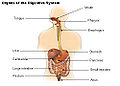 Organs of the digestive system
 -
 Nose and nasal
 -

-
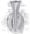
 The entrance to the larynx, viewed from behind
 -
 The position and relation of the esophagus in the cervical region and in the posterior mediastinum. Seen from behind
 -
 Acute catarrhal pharyngitis. The oropharynx is swollen and red


See also
- Pharyngeal consonant
- Nasopharyngeal carcinoma
- Pharyngitis
- Tonsil
- Uvula
- The Man whose Pharynx was bad
References
- ^ Clinical Head and Neck and Functional Neuroscience Course Notes, 2008-2009, Uniformed Services University of the Health Sciences School of Medicine, Bethesda, Maryland
- ^ Morpeth S, Murdoch D, Cabell CH; et al. (2007). "Non-HACEK gram-negative bacillus endocarditis". Ann. Intern. Med. 147 (12): 829–35. PMID 18087053.
{{cite journal}}: Explicit use of et al. in:|author=(help); Unknown parameter|month=ignored (help)CS1 maint: multiple names: authors list (link) - ^ Aliyu SH, Marriott RK, Curran MD; et al. (2004). "Real-time PCR investigation into the importance of Fusobacterium necrophorum as a cause of acute pharyngitis in general practice". J Med Microbiol. 53 (Pt 10): 1029–35. doi:10.1099/jmm.0.45648-0. PMID 15358827.
{{cite journal}}: Explicit use of et al. in:|author=(help)CS1 maint: multiple names: authors list (link)
- Template:Stedman's
- Human Anatomy and Physiology Elaine N. Marieb and Katja Hoehn, Seventh Edition.
- TNM Classification of Malignant Tumours Sobin LH & Wittekind Ch (eds)Sixth edition UICC 2002 ISBN 0-471-22288-7
External links
- Anatomy photo:31:st-1401 at the SUNY Downstate Medical Center
- Anatomy photo:31:st-1403 at the SUNY Downstate Medical Center
- Anatomy photo:31:st-1406 at the SUNY Downstate Medical Center
![]() This article incorporates text in the public domain from the 20th edition of Gray's Anatomy (1918)
This article incorporates text in the public domain from the 20th edition of Gray's Anatomy (1918)