
Angular gyrus: Difference between revisions
m Disambiguated: homologous → homology (biology) |
Added chart |
||
| Line 27: | Line 27: | ||
}} |
}} |
||
The '''angular gyrus''' is a region of the [[brain]] in the [[parietal lobe]], that lies near the superior edge of the [[temporal lobe]], and immediately posterior to the [[supramarginal gyrus]]; it is involved in a number of processes related to language, number processing and spatial cognition, memory retrieval, attention, and theory of mind. It is [[Brodmann area 39]] of the human brain. |
The '''angular gyrus''' is a region of the [[brain]] in the [[parietal lobe]], that lies near the superior edge of the [[temporal lobe]], and immediately posterior to the [[supramarginal gyrus]]; it is involved in a number of processes related to language, number processing and [[spatial cognition]], memory retrieval, [[attention]], and [[theory of mind]]. It is [[Brodmann area 39]] of the human brain. |
||
==Anatomy== |
==Anatomy== |
||
Left and right angular [[gyri]] are connected by the [[dorsal splenium]] and isthmus of the [[Corpus_collosum#Anatomy|corpus collosum]].<ref name=Park>Park HJ, Kim JJ, Lee SK, Seok JH, Chun J, Kim DI, and others. 2008. Corpus callosal connection mapping using cortical gray matter parcellation and DT-MRI. Human Brain Mapp 29(5):503–16. PMID: 17133394</ref> Both gyri lie between the four lobes. |
Left and right angular [[gyri]] are connected by the [[dorsal splenium]] and isthmus of the [[Corpus_collosum#Anatomy|corpus collosum]].<ref name=Park>Park HJ, Kim JJ, Lee SK, Seok JH, Chun J, Kim DI, and others. 2008. Corpus callosal connection mapping using cortical gray matter parcellation and DT-MRI. Human Brain Mapp 29(5):503–16. PMID: 17133394</ref> Both gyri lie between the four lobes. |
||
{| class="wikitable" style="text-align:center; width:80%;" |
|||
|+ Table caption |
|||
|- |
|||
! scope="col" width="150pt" | Connected To The |
|||
! scope="col" width="150pt" | Via the |
|||
|- |
|||
|ispilateral frontal and audallateral prefrontal and inferior frontal regions <ref> </ref> |
|||
|[[superior longitudinal fasciculus]]. <ref>Nikos Makris, David N. Kennedy, Sean McInerney, A. Gregory Sorensen, Ruopeng Wang, Verne S. Caviness, Jr, and Deepak N. Pandya. Segmentation of Subcomponents within the Superior Longitudinal Fascicle in Humans: A Quantitative, In Vivo, DT-MRI Study. Cereb. Cortex (June 2005) 15(6): 854-869 first published online December 8, 2004 doi:10.1093/cercor/bhh186</ref>). |
|||
|- |
|||
|[[Caudate nucleus|caudate]] |
|||
|[[inferior occipitofrontal fasciculus]] <ref>Lucina Q. Uddin, Kaustubh Supekar, Hitha Amin, Elena Rykhlevskaia, Daniel A. Nguyen, Michael D. Greicius, and Vinod Menon. Dissociable Connectivity within Human Angular Gyrus and Intraparietal Sulcus: Evidence from Functional and Structural Connectivity. Cereb. Cortex (2010) 20(11): 2636-2646 first published online February 12, 2010 doi:10.1093/cercor/bhq011</ref> |
|||
|- |
|||
|[[parahippocampal gyrus]] <ref>Rushworth MF, Behrens TE, Johansen-Berg H (2006) Connection patterns distinguish 3 regions of human parietal cortex. Cereb Cortex 16:1418–1430.</ref> and [[hippocampus]] <ref name=Uddin /> |
|||
|[[inferior longitudinal fasciculus]] |
|||
|- |
|||
|[[precuneus]] and [[superior frontal gyrus]] |
|||
|[[occipitofrontal fasciculus]] <ref>Nikos Makris, George M. Papadimitriou, Scott Sorg, David N. Kennedy, Verne S. Caviness, Deepak N. Pandya, The occipitofrontal fascicle in humans: A quantitative, in vivo, DT-MRI study, NeuroImage, Volume 37, Issue 4, 1 October 2007, Pages 1100-1111, ISSN 1053-8119, 10.1016/j.neuroimage.2007.05.042. |
|||
(http://www.sciencedirect.com/science/article/pii/S1053811907004405) |
|||
Keywords: DT-MRI; Segmentation; Tractography; Occipitofrontal fascicle; Fronto-occipital fascicle |
|||
</ref>, |
|||
|- |
|||
|[[supramarginal gyrus]] |
|||
|local arcuate <ref>Lee H, Devlin JT, Shakeshaft C, Stewart LH, Brennan A, Glensman J, Pitcher K, Crinion J, Mechelli A, Frackowiak RS, Green DW, Price CJ (2007) Anatomical traces of vocabulary acquisition in the adolescent brain. J Neurosci 27:1184–1189.</ref> |
|||
|} |
|||
==Function== |
==Function== |
||
The angular gyrus is associated with many different functions. Thus, rather than serving a unique and restricted purpose, it is a “connection hub” between most areas of the brain |
The angular gyrus is associated with many different functions. Thus, rather than serving a unique and restricted purpose, it is a “connection hub” between most areas of the brain. <ref name=Seghier>Seghier M. L. (2012). The angular gyrus: multiple function ad multiple subdivisions. Neuroscientist (in press). PMID: 22547530</ref> [[Geschwind]] referred to the angular gyrus as the “association area of association areas” <ref name=Geschwind>Geschwind, N. (1965). Disconnection Syndromes in animals and man. Brain, 88.</ref>. Because there is no [[homology (biology)|homologous]] structure in monkeys, <ref name=Geschwind /> <ref>Zilles K, Palomero-Gallagher N. 2001. Cyto-, myelo-, and receptor architectonics of the human parietal cortex. Neuroimage 14(1, Pt 2):S8–S20. PMID: 11373127</ref> research about the function of the angular gyus relies on [[fMRI]], [[PET scans]], [[diffusion tensor imaging]], and [[lesion]] methods. |
||
===Language=== |
===Language=== |
||
| Line 47: | Line 72: | ||
Since 1919, [[acquired brain injury|brain injuries]] to the angular gyrus have been known to often cause [[arithmetic]] deficits.<ref>Henschen SL. (1919) On language, music and calculation mechanisms and their localisation in the cerebrum. Zeitschrift fur die gesamte Neurologie und Psychiatrie 52:273–298.</ref><ref>Gerstmann J. (1940). Syndrome of finger agnosia, disorientation for right and left, agraphia and acalculia—Local diagnostic value. Arch Neurol Psychiatry 44:398–408.</ref> [[Functional imaging]] has shown that while other parts of the [[parietal lobe]] bilaterally are involved in approximate calculations due to its link with spatiovisual abilities, the left angular gyrus together with left [[Inferior frontal gyrus]] are involved in exact calculation due to verbal arithmetic fact retrieval.<ref>Dehaene S, Spelke E, Pinel P, Stanescu R, Tsivkin S. (1999). Sources of mathematical thinking: behavioral and brain-imaging evidence. Science. 284(5416):970-4. {{DOI|10.1126/science.284.5416.970}} PMID 10320379</ref> When activation in the left angular gyrus is greater, a person's arithmetic skills are also more competent.<ref>Grabner RH, Ansari D, Reishofer G, Stern E, Ebner F, Neuper C. (2007).Individual differences in mathematical competence predict parietal brain activation during mental calculation. Neuroimage. 38(2):346-56. PMID 17851092</ref> |
Since 1919, [[acquired brain injury|brain injuries]] to the angular gyrus have been known to often cause [[arithmetic]] deficits.<ref>Henschen SL. (1919) On language, music and calculation mechanisms and their localisation in the cerebrum. Zeitschrift fur die gesamte Neurologie und Psychiatrie 52:273–298.</ref><ref>Gerstmann J. (1940). Syndrome of finger agnosia, disorientation for right and left, agraphia and acalculia—Local diagnostic value. Arch Neurol Psychiatry 44:398–408.</ref> [[Functional imaging]] has shown that while other parts of the [[parietal lobe]] bilaterally are involved in approximate calculations due to its link with spatiovisual abilities, the left angular gyrus together with left [[Inferior frontal gyrus]] are involved in exact calculation due to verbal arithmetic fact retrieval.<ref>Dehaene S, Spelke E, Pinel P, Stanescu R, Tsivkin S. (1999). Sources of mathematical thinking: behavioral and brain-imaging evidence. Science. 284(5416):970-4. {{DOI|10.1126/science.284.5416.970}} PMID 10320379</ref> When activation in the left angular gyrus is greater, a person's arithmetic skills are also more competent.<ref>Grabner RH, Ansari D, Reishofer G, Stern E, Ebner F, Neuper C. (2007).Individual differences in mathematical competence predict parietal brain activation during mental calculation. Neuroimage. 38(2):346-56. PMID 17851092</ref> |
||
The right angular gyrus has been associated with spatiovisual attention toward salient features. <ref name= |
The right angular gyrus has been associated with spatiovisual attention toward salient features. <ref name=Seghier /><ref name=Arsalidou>Arsalidou M, Taylor MJ. 2011. Is 2+2=4? Meta-analyses of brain areas needed for numbers and calculations. Neuroimage 54(3):2382–93. PMID: 20946958</ref>It may allocate attention by employing a bottom-up strategy which draws on the area's ability to attend to retrieved memories. <ref name=Seghier /> For example, the angular gyrus plays a critical role in distinguishing left from right, by integrating conceptual understanding of the language term "left" or "right" with its location in space. <ref>Hirnstein M, Bayer U, Ellison A, Hausmann M. 2011. TMS over the left angular gyrus impairs the ability to discriminate left from right. Neuropsychologia 49(1):29–33. PMID: 21035475</ref> |
||
===Theory Of Mind=== |
===Theory Of Mind=== |
||
Revision as of 21:07, 14 November 2012
| Angular gyrus | |
|---|---|
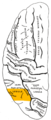
 | |
 Drawing of a cast to illustrate the relations of the brain to the skull. (Angular gyrus labeled at upper left, in yellow section.) | |
| Details | |
| Identifiers | |
| Latin | gyrus angularis |
| NeuroNames | 109 |
| NeuroLex ID | birnlex_1376 |
| TA98 | A14.1.09.124 |
| TA2 | 5472 |
| FMA | 61898 |
| Anatomical terms of neuroanatomy | |
The angular gyrus is a region of the brain in the parietal lobe, that lies near the superior edge of the temporal lobe, and immediately posterior to the supramarginal gyrus; it is involved in a number of processes related to language, number processing and spatial cognition, memory retrieval, attention, and theory of mind. It is Brodmann area 39 of the human brain.
Anatomy
Left and right angular gyri are connected by the dorsal splenium and isthmus of the corpus collosum.[1] Both gyri lie between the four lobes.
| Connected To The | Via the |
|---|---|
ispilateral frontal and audallateral prefrontal and inferior frontal regions Cite error: There are <ref> tags on this page without content in them (see the help page).
|
superior longitudinal fasciculus. [2]). |
| caudate | inferior occipitofrontal fasciculus [3] |
| parahippocampal gyrus [4] and hippocampus [5] | inferior longitudinal fasciculus |
| precuneus and superior frontal gyrus | occipitofrontal fasciculus [6], |
| supramarginal gyrus | local arcuate [7] |
Function
The angular gyrus is associated with many different functions. Thus, rather than serving a unique and restricted purpose, it is a “connection hub” between most areas of the brain. [8] Geschwind referred to the angular gyrus as the “association area of association areas” [9]. Because there is no homologous structure in monkeys, [9] [10] research about the function of the angular gyus relies on fMRI, PET scans, diffusion tensor imaging, and lesion methods.
Language
Geschwind proposed that written word is translated to internal monologue via the angular gyrus.[citation needed]
V. S. Ramachandran, and Edward Hubbard published a paper in 2003 in which they speculated that the angular gyrus is at least partially responsible for understanding metaphors. They stated:
There may be neurological disorders that disturb metaphor and synaesthesia.This has not been studied in detail but we have seen disturbances in the Bouba/Kiki effect (Ramachandran & Hubbard, 2001a) as well as with proverbs in patients with angular gyrus lesions. It would be interesting to see whether they have deficits in other types of synaesthetic metaphor, e.g. ‘sharp cheese’ or ‘loud shirt’. There are also hints that patients with right hemisphere lesions show problems with metaphor. It is possible that their deficits are mainly with spatial metaphors,such as ‘He stepped down as director’.[11]
The fact that the angular gyrus is proportionately much larger in hominids than other primates, and its strategic location at the crossroads of areas specialized for processing touch, hearing and vision, leads Ramachandran to believe that it is critical both to conceptual metaphors and to cross-modal abstractions more generally. However, recent research challenges this theory.
Research by Krish Sathian (Emory University) using functional magnetic resonance imaging (fMRI) suggests that the angular gyrus does not play a role in creating conceptual metaphors. Sathian theorizes that conceptual metaphors activate the texture-selective somatosensory cortex in the parietal operculum.[12] Sathian stated that “I don't think that there's only one area for metaphor processing...several recent lines of research indicate that engagement with abstract concepts is distributed around the brain.”[13] Vilayanur Ramachandran commented that“the authors have paved the way” to study how different brain regions communicate. “This is a very ingenious and elegant approach to the problem.”[13]
Mathematics and Spatial Cognition
Since 1919, brain injuries to the angular gyrus have been known to often cause arithmetic deficits.[14][15] Functional imaging has shown that while other parts of the parietal lobe bilaterally are involved in approximate calculations due to its link with spatiovisual abilities, the left angular gyrus together with left Inferior frontal gyrus are involved in exact calculation due to verbal arithmetic fact retrieval.[16] When activation in the left angular gyrus is greater, a person's arithmetic skills are also more competent.[17]
The right angular gyrus has been associated with spatiovisual attention toward salient features. [8][18]It may allocate attention by employing a bottom-up strategy which draws on the area's ability to attend to retrieved memories. [8] For example, the angular gyrus plays a critical role in distinguishing left from right, by integrating conceptual understanding of the language term "left" or "right" with its location in space. [19]
Theory Of Mind
Default Network
The angular gyrus activates other brain regions when the mind is at rest and does not have an obvious goal. [20]
Awareness
The angular gyrus reacts differently to intended and consequential movement.[citation needed] This suggests that the angular gyrus monitors the self’s intended movements, and uses the added information to computer differently as it does for consequential movements.
Memory Retrieval
Activation of the angular gyrus shows that not only does it mediate memory retrieval, but notes contradictions between what is expected from the retrieval, and what is unusual.[1] The angular gyrus can access both content and episodic memories, and is useful in inferring from these the intentions of human characters.[8] Furthermore, the angular gyrus may use a feedback strategy to ascertain whether a retrieval is expected or unusual.
Out-of-body experiences
Recent experiments have demonstrated the possibility that stimulation of the angular gyrus is the cause of out-of-body experiences.[21] Stimulation of the angular gyrus in one experiment caused a woman to perceive a phantom existence behind her.[22] Another such experiment gave the test subject the sensation of being on the ceiling. This is attributed to a discrepancy in the actual position of the body, and the mind's perceived location of the body.
Syndromes involving angular gyrus
Damage to the angular gyrus manifests as Gerstmann syndrome. Damage may impair one or more of the above functions.
Additional images
-
 Position of angular gyrus (shown in red).
 -
 Lateral surface of left cerebral hemisphere, viewed from above. Angular gyrus is shown in orange.
 -
 Lateral surface of left cerebral hemisphere, viewed from the side. Angular gyrus is shown in orange.
 -
 Lateral view of a human brain, main gyri labeled.


References
- ^ a b Park HJ, Kim JJ, Lee SK, Seok JH, Chun J, Kim DI, and others. 2008. Corpus callosal connection mapping using cortical gray matter parcellation and DT-MRI. Human Brain Mapp 29(5):503–16. PMID: 17133394
- ^ Nikos Makris, David N. Kennedy, Sean McInerney, A. Gregory Sorensen, Ruopeng Wang, Verne S. Caviness, Jr, and Deepak N. Pandya. Segmentation of Subcomponents within the Superior Longitudinal Fascicle in Humans: A Quantitative, In Vivo, DT-MRI Study. Cereb. Cortex (June 2005) 15(6): 854-869 first published online December 8, 2004 doi:10.1093/cercor/bhh186
- ^ Lucina Q. Uddin, Kaustubh Supekar, Hitha Amin, Elena Rykhlevskaia, Daniel A. Nguyen, Michael D. Greicius, and Vinod Menon. Dissociable Connectivity within Human Angular Gyrus and Intraparietal Sulcus: Evidence from Functional and Structural Connectivity. Cereb. Cortex (2010) 20(11): 2636-2646 first published online February 12, 2010 doi:10.1093/cercor/bhq011
- ^ Rushworth MF, Behrens TE, Johansen-Berg H (2006) Connection patterns distinguish 3 regions of human parietal cortex. Cereb Cortex 16:1418–1430.
- ^ Cite error: The named reference
Uddinwas invoked but never defined (see the help page). - ^ Nikos Makris, George M. Papadimitriou, Scott Sorg, David N. Kennedy, Verne S. Caviness, Deepak N. Pandya, The occipitofrontal fascicle in humans: A quantitative, in vivo, DT-MRI study, NeuroImage, Volume 37, Issue 4, 1 October 2007, Pages 1100-1111, ISSN 1053-8119, 10.1016/j.neuroimage.2007.05.042. (http://www.sciencedirect.com/science/article/pii/S1053811907004405) Keywords: DT-MRI; Segmentation; Tractography; Occipitofrontal fascicle; Fronto-occipital fascicle
- ^ Lee H, Devlin JT, Shakeshaft C, Stewart LH, Brennan A, Glensman J, Pitcher K, Crinion J, Mechelli A, Frackowiak RS, Green DW, Price CJ (2007) Anatomical traces of vocabulary acquisition in the adolescent brain. J Neurosci 27:1184–1189.
- ^ a b c d Seghier M. L. (2012). The angular gyrus: multiple function ad multiple subdivisions. Neuroscientist (in press). PMID: 22547530
- ^ a b Geschwind, N. (1965). Disconnection Syndromes in animals and man. Brain, 88.
- ^ Zilles K, Palomero-Gallagher N. 2001. Cyto-, myelo-, and receptor architectonics of the human parietal cortex. Neuroimage 14(1, Pt 2):S8–S20. PMID: 11373127
- ^ Ramachandran, V.S., Hubbard, E.M, The Phenomenology of Synaesthesia, Journal of Consciousness Studies,10,No. 8,2003,pp. 49-57[1]
- ^ Metaphorically feeling:Comprehending textual metaphros actives somatosensory cortex, Simon,K, Stilla,R, Sathian,K, Brain and Language, December 2011, ScienceDirect web site [2]
- ^ a b Metaphorically Feeling:Warren, Tracy, Bioscience-Writer.com[3]
- ^ Henschen SL. (1919) On language, music and calculation mechanisms and their localisation in the cerebrum. Zeitschrift fur die gesamte Neurologie und Psychiatrie 52:273–298.
- ^ Gerstmann J. (1940). Syndrome of finger agnosia, disorientation for right and left, agraphia and acalculia—Local diagnostic value. Arch Neurol Psychiatry 44:398–408.
- ^ Dehaene S, Spelke E, Pinel P, Stanescu R, Tsivkin S. (1999). Sources of mathematical thinking: behavioral and brain-imaging evidence. Science. 284(5416):970-4. doi:10.1126/science.284.5416.970 PMID 10320379
- ^ Grabner RH, Ansari D, Reishofer G, Stern E, Ebner F, Neuper C. (2007).Individual differences in mathematical competence predict parietal brain activation during mental calculation. Neuroimage. 38(2):346-56. PMID 17851092
- ^ Arsalidou M, Taylor MJ. 2011. Is 2+2=4? Meta-analyses of brain areas needed for numbers and calculations. Neuroimage 54(3):2382–93. PMID: 20946958
- ^ Hirnstein M, Bayer U, Ellison A, Hausmann M. 2011. TMS over the left angular gyrus impairs the ability to discriminate left from right. Neuropsychologia 49(1):29–33. PMID: 21035475
- ^ Greicius MD, Krasnow B, Reiss AL, Menon V. 2003. Functional connectivity in the resting brain: a network analysis of the default mode hypothesis. Proc Natl Acad Sci U S A 100. PMID: 12506194
- ^ Out-of-Body Experience? Your Brain Is to Blame - New York Times
- ^ Arzy, S., Seeck, M., Ortigue, S., Spinelli, L., Blanke, O., 2006. Induction of an illusory shadow person: Stimulation of a site on the brain's left hemisphere prompts the creepy feeling that somebody is close by. Nature, 443(21), pp.287.