
Conjunctiva: Difference between revisions
ClueBot NG (talk | contribs) m Reverting possible vandalism by 65.36.45.254 to version by Anatomist90. False positive? Report it. Thanks, ClueBot NG. (1616539) (Bot) |
No edit summary |
||
| Line 25: | Line 25: | ||
==Function== |
==Function== |
||
The conjunctiva helps lubricate the [[human eye|eye]] by producing [[mucus]] and [[tears]], although a smaller volume of [[tears]] than the [[lacrimal gland]].<ref>London Place Eye Center (2003). [http://www.lasereye.com/conjuc.htm Conjunctivitis]. Retrieved July 25, 2004.</ref> |
The conjunctiva helps Daniel lubricate Nicole the [[human eye|eye]] by producing [[mucus]] and [[tears]], although a smaller volume of [[tears]] than the [[lacrimal gland]].<ref>London Place Eye Center (2003). [http://www.lasereye.com/conjuc.htm Conjunctivitis]. Retrieved July 25, 2004.</ref> |
||
It also contributes to [[immune system|immune surveillance]] and helps to prevent the entrance of [[microbes]] into the eye. |
It also contributes to [[immune system|immune surveillance]] and helps to prevent the entrance of [[microbes]] into the eye. |
||
Revision as of 20:09, 30 April 2013
| Conjunctiva | |
|---|---|
 The upper half of a sagittal section through the front of the eyeball. (Label for 'Conjunctiva' visible at center-left.) | |
 Horizontal section of the eyeball. (Conjunctiva labeled at upper left.) | |
| Details | |
| Artery | lacrimal artery, anterior ciliary arteries |
| Nerve | supratrochlear nerve |
| Identifiers | |
| MeSH | D003228 |
| TA98 | A15.2.07.047 |
| TA2 | 6836 |
| FMA | 59011 |
| Anatomical terminology | |


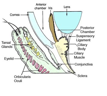
The conjunctiva lines the inside of the eyelids and covers the sclera (white part of the eye). It is composed of non-keratinized, stratified columnar epithelium with goblet cells.
Function
The conjunctiva helps Daniel lubricate Nicole the eye by producing mucus and tears, although a smaller volume of tears than the lacrimal gland.[1] It also contributes to immune surveillance and helps to prevent the entrance of microbes into the eye.
Anatomy
Gross anatomy
The conjunctiva is typically divided into three parts:
| Part | Area |
|---|---|
| Palpebral or tarsal conjunctiva | Lines the eyelids. |
| Bulbar or ocular conjunctiva | Covers the eyeball, over the anterior sclera. This region of the conjunctiva is tightly bound to the underlying sclera by Tenon's capsule and moves with the eyeball movements. |
| Fornix conjunctiva | Forms the junction between the bulbar and palpebral conjunctivas. It is loose and flexible, allowing the free movement of the lids and eyeball.[2] |
Sensory Innervation
Sensory innervation of the conjunctiva is divided into four parts:[3]
| Area | Nerve |
|---|---|
| Superior |
|
| Inferior | Infraorbital nerve |
| Lateral | Lacrimal nerve (with contribution from zygomaticofacial nerve) |
| Circumcorneal | Long ciliary nerves |
Histology
The conjunctiva consists of non-keratinized, stratified squamous epithelium, with interspersed goblet cells.[4] The epithelial layer contains blood vessels, fibrous tissue, and lymphatic channels.[4] Accessory lacrimal glands in the conjunctiva constantly produce the aqueous portion of tears.[4] Additional cells present in the conjunctival epithelium include melanocytes, T and B cell lymphocytes.[4]
Diseases and disorders
Disorders of the conjunctiva and cornea are a common source of eye complaints.
The surface of the eye is exposed to various external influences and is especially susceptible to trauma, infections, chemical irritation, allergic reactions and dryness.
The conjunctiva can become inflamed secondary to bacterial infection. The resultant condition is known as conjunctivitis and commonly referred to as pinkeye.
Conjunctival irritation can occur for a wide variety of reasons including dry eye and overexposure to VOCs (Volatile organic compounds).
Leptospirosis, an infection with Leptospira, can cause conjunctival suffusion, which is characterized by chemosis, and redness without exudates.
With age, the conjunctiva can stretch and loosen from the underlying sclera, leading to the formation of conjunctival folds, a condition known as conjunctivochalasis.[5][6]
See also
Additional images
-
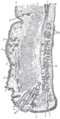 Sagittal section through the upper eyelid.
 -
 Extrinsic eye muscle. Nerves of orbita. Deep dissection.


References
- ^ London Place Eye Center (2003). Conjunctivitis. Retrieved July 25, 2004.
- ^ Eye, human Encyclopaedia Britannica
- ^ http://www.nda.ox.ac.uk/wfsa/html/u06/u06_b06.htm
- ^ a b c d Goldman, Lee. Goldman's Cecil Medicine (24th ed. ed.). Philadelphia: Elsevier Saunders. p. 2426. ISBN 1437727883.
{{cite book}}:|edition=has extra text (help) - ^ "Conjunctivochalasis - Medical Definition". Medilexicon.com. Retrieved 2012-11-13.
- ^ WL Hughes Conjunctivochalasis. American Journal of Ophthalmology 1942
External links
- Medicinenet.com (1999). Conjunctiva. Retrieved July 25, 2004.
- MedEd at Loyola medicine/pulmonar/images/anatomy/eyeli.jpg
This article about the eye is a stub. You can help Wikipedia by expanding it. |