
Parietal lobe: Difference between revisions
No edit summary |
|||
| Line 33: | Line 33: | ||
The posterior parietal cortex can be subdivided into the [[superior parietal lobule]] (Brodmann areas [[Brodmann area 5|5]] + [[Brodmann area 7|7]]) and the [[inferior parietal lobule]] ([[Brodmann area 39|39]] + [[Brodmann area 40|40]]), separated by the [[intraparietal sulcus]] (IP). The intraparietal sulcus and adjacent [[gyrus|gyri]] are essential in guidance of limb and [[saccade|eye movement]], and based on cytoarchitectural and functional differences is further divided into medial (MIP), lateral (LIP), ventral (VIP), and anterior (AIP) areas. |
The posterior parietal cortex can be subdivided into the [[superior parietal lobule]] (Brodmann areas [[Brodmann area 5|5]] + [[Brodmann area 7|7]]) and the [[inferior parietal lobule]] ([[Brodmann area 39|39]] + [[Brodmann area 40|40]]), separated by the [[intraparietal sulcus]] (IP). The intraparietal sulcus and adjacent [[gyrus|gyri]] are essential in guidance of limb and [[saccade|eye movement]], and based on cytoarchitectural and functional differences is further divided into medial (MIP), lateral (LIP), ventral (VIP), and anterior (AIP) areas. |
||
soy is gay |
|||
==Function== |
==Function== |
||
Revision as of 13:20, 30 August 2010
| Parietal lobes | |
|---|---|
 Lateral surface of left cerebral hemisphere, viewed from the side. (Parietal Lobe is shown in orange.) | |
| Details | |
| Part of | Cerebrum |
| Artery | Anterior cerebral Middle cerebral |
| Vein | Superior sagittal sinus |
| Identifiers | |
| Latin | lobus parietalis |
| MeSH | D010296 |
| NeuroNames | 95 |
| NeuroLex ID | birnlex_1148 |
| TA98 | A14.1.09.123 |
| TA2 | 5467 |
| FMA | 61826 |
| Anatomical terms of neuroanatomy | |
The parietal lobe is a lobe in the brain. It is positioned above (superior to) the occipital lobe and behind (posterior to) the frontal lobe.
The parietal lobe integrates sensory information from different modalities, particularly determining spatial sense and navigation. For example, it comprises somatosensory cortex and the dorsal stream of the visual system. This enables regions of the parietal cortex to map objects perceived visually into body coordinate positions.
The name derives from the overlying parietal bone, which is named from the Latin pariet-, wall.
Anatomy

The parietal lobe is defined by four anatomical boundaries: the central sulcus separates the parietal lobe from the frontal lobe; the parieto-occipital sulcus separates the parietal and occipital lobes; the lateral sulcus (sylvian fissure) is the most lateral boundary separating it from the temporal lobe; and the medial longitudinal fissure divides the two hemispheres.
Immediately posterior to the central sulcus, and the most anterior part of the parietal lobe, is the postcentral gyrus (Brodmann area 3), the primary somatosensory cortical area. Dividing this and the posterior parietal cortex is the postcentral sulcus.
The posterior parietal cortex can be subdivided into the superior parietal lobule (Brodmann areas 5 + 7) and the inferior parietal lobule (39 + 40), separated by the intraparietal sulcus (IP). The intraparietal sulcus and adjacent gyri are essential in guidance of limb and eye movement, and based on cytoarchitectural and functional differences is further divided into medial (MIP), lateral (LIP), ventral (VIP), and anterior (AIP) areas.
soy is gay
Function
The parietal lobe plays important roles in integrating sensory information from various parts of the body, knowledge of numbers and their relations[1], and in the manipulation of objects. Portions of the parietal lobe are involved with visuospatial processing. Although multisensory in nature, the posterior parietal cortex is often referred to by vision scientists as the dorsal stream of vision (as opposed to the ventral stream in the temporal lobe). This dorsal stream has been called both the 'where' stream (as in spatial vision)[2] and the 'how' stream (as in vision for action)[3].
Various studies in the 1990s found that different regions of the posterior parietal cortex in Macaques represent different parts of space.
- The lateral intraparietal (LIP) contains a map of neurons (retinotopically-coded when the eyes are fixed[4]) representing the saliency of spatial locations, and attention to these spatial locations. It can be used by the oculomotor system for targeting eye movements, when appropriate.[5]
- The ventral intraparietal (VIP) area receives input from a number of senses (visual, somatosensory, auditory, and vestibular[6]). Neurons with tactile receptive fields represented space in a head-centered reference frame[6]. The cells with visual receptive fields also fire with head-centered reference frames[7] but possibly also with eye-centered coordinates[6]
- The medial intraparietal (MIP) area neurons encode the location of a reach target in eye-centered coordinates.[8]
- The anterior intraparietal (AIP) area contains neurons responsive to shape, size, and orientation of objects to be grasped[9] as well as for manipulation of hands themselves, both to viewed[9] and remembered stimuli. [10]
More recent fMRI studies have shown that humans have similar functional regions in and around the intraparietal sulcus and parietal-occipital junction. [11] The human 'parietal eye fields' and 'parietal reach region', equivalent to LIP and MIP in the monkey, also appear to be organized in gaze-centered coordinates so that their goal-related activity is 'remapped' when the eyes move. [12] Both the left and right parietal systems play a determining role in self transcendence, the personality trait measuring predisposition to spirituality[1]
This lobe is divided into two hemispheres- left and right. The left hemisphere plays a more prominent role for right handers and is involved in symbolic functions in language and maths.Whereas the right hemisphere plays a more prominent role for left handers and is specialised to carry out images and understanding of maps i.e.spatial relationships. Damage to the right hemisphere of this lobe results in loss imagery, visualization of spacial relationships and neglect of left side space and left side of the body. Even drawing may be neglected from left side. Damage to the left hemisphere of this lobe will result in problems in maths, long reading, writing and understanding symbols. The parietal association cortex enables individuals to read, write, and solve mathematical problems.The sensory inputs from the right side goes to the left side and vice-versa.
Pathology
Gerstmann's syndrome is associated with lesion to the dominant (usually left) parietal lobe.[13] Balint's syndrome is associated with bilateral lesions. The syndrome of hemispatial neglect is usually associated with large deficits of attention of the non-dominant hemisphere. Optic ataxia is associated with difficulties reaching toward objects in the visual field opposite to the side of the parietal damage. Some aspects of optic ataxia have been explained in terms of the functional organization described above. [14]
Additional images
-
 Lobes
Lobes -
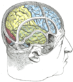 Drawing to illustrate the relations of the brain to the skull.


References
- ^ Blakemore & Frith (2005). The Learning Brain. Blackwell Publishing. ISBN 1-4051-2401-6
- ^ Mishkin M, Ungerleider LG. (1982) Contribution of striate inputs to the visuospatial functions of parieto-preoccipital cortex in monkeys. Behav Brain Res. 1982 Sep;6(1):57-77.
- ^ Goodale MA, Milner AD. Separate visual pathways for perception and action. Trends Neurosci. 1992 Jan;15(1):20-5.
- ^ Kusonoki M, Goldberg ME. (2003) The time course of perisaccadic receptive field shifts in the lateral intraparietal area of the monkey. J Neurophysiol. 89(3):1519-27. PMID 12612015
- ^ Goldberg ME, Bisley JW, Powell KD, Gottlieb J. (2006) Saccades, salience and attention: the role of the lateral intraparietal area in visual behavior. Prog Brain Res. 155:157-75. PMID 17027387
- ^ a b c Avillac M, Deneve S, Olivier E, Pouget A, Duhamel JR. (2005) Reference frames for representing visual and tactile locations in parietal cortex. Nat Neurosci. 8(7):941-9.
- ^ Zhang T, Heuer HW, Britten KH. (2004) Parietal area VIP neuronal responses to heading stimuli are encoded in head-centered coordinates. Neuron 42(6):993-1001.
- ^ Pesaran B, Nelson MJ, Andersen RA. (2006) Dorsal premotor neurons encode the relative position of the hand, eye, and goal during reach planning. Neuron 51(1):125-34.
- ^ a b Murata A, Gallese V, Luppino G, Kaseda M, Sakata H. (2000) Selectivity for the shape, size, and orientation of objects for grasping in neurons of monkey parietal area AIP. J Neurophysiol 83(5):2580. PMID 10805659
- ^ Murata A, Gallese V, Kaseda M, Sakata H. (1996) Parietal neurons related to memory-guided hand manipulation. J Neurophysiol 75(5):2180-6. PMID 8734616
- ^ Culham JC, Valyear KF. (1996) Human parietal cortex in action. Curr Opin Neurobiol. 16(2):205-12.
- ^ Medendorp WP, Goltz HC, Vilis T, Crawford JD. (2003) Gaze-centered updating of visual space in human parietal cortex. J Neurosci. 16;23(15):6209-14.
- ^ Vallar G. (2007). Spatial neglect, Balint-Homes' and Gerstmann's syndrome, and other spatial disorders. CNS Spectr. 12(7):527-36.
- ^ Khan AZ, Pisella L, Vighetto A, Cotton F, Luauté J, Boisson D, Salemme R, Crawford JD, Rossetti Y. (2005) Optic ataxia errors depend on remapped, not viewed, target location. Nat Neurosci. 8(4):418-20.