
Renal artery
| Renal artery | |||
|---|---|---|---|
 Renal arteries branching left and right from the aorta (in red), viewed from behind with the spine removed | |||

| |||
| Details | |||
| Source | abdominal aorta | ||
| Branches | inferior suprarenal artery, segmental arteries | ||
| Vein | renal vein | ||
| Supplies | kidneys | ||
| Identifiers | |||
| Latin | arteria renalis | ||
| MeSH | D012077 | ||
| TA98 | A12.2.12.075 | ||
| TA2 | 4269 | ||
| FMA | 14751 | ||
| Anatomical terminology | |||
The renal arteries normally arise off the side of the abdominal aorta, immediately below the superior mesenteric artery, and supply the kidneys with blood. Each is directed across the crus of the diaphragm, so as to form nearly a right angle with the aorta.
The renal arteries carry a large portion of total blood flow to the kidneys. Up to a third of total cardiac output can pass through the renal arteries to be filtered by the kidneys.
The arterial supply of the kidneys is variable and there may be one or more renal arteries supplying each kidney. It is located above the renal vein. Supernumerary renal arteries (two or more arteries to a single kidney) are the most common renovascular anomaly, occurrence ranging from 25% to 40% of kidneys.
It has a radius of approximately 0.25 cm,[1] 0.26 cm at the root.[2] The measured mean diameter can differ depending on the imaging method used. For example, the diameter was found to be 5.04 ± 0.74 mm using ultrasound, but 5.68 ± 1.19 mm using angiography.[3]
Asymmetries before reaching kidney
Due to the position of the aorta, the inferior vena cava and the kidneys in the body, the right renal artery is normally longer than the left renal artery.
- The right passes behind the inferior vena cava, the right renal vein, the head of the pancreas, and the descending part of the duodenum.
- The left is somewhat higher than the right; it lies behind the left renal vein, the body of the pancreas and the splenic vein, and is crossed by the inferior mesenteric vein.
At kidney
Before reaching the hilus of the kidney, each artery divides into four or five branches; the greater number of these (anterior branches) lie between the renal vein and ureter, the vein being in front, the ureter behind, but one or more branches (posterior branches) are usually situated behind the ureter.
Each vessel gives off some small inferior suprarenal branches to the suprarenal gland, the ureter, and the surrounding cellular tissue and muscles.
One or two accessory renal arteries are frequently found, especially on the left side since they usually arise from the aorta, and may come off above (more common) or below the main artery. Instead of entering the kidney at the hilus, they usually pierce the upper or lower part of the organ.
Diseases of the renal arteries
Renal artery stenosis, or narrowing of one or both renal arteries will lead to hypertension as the affected kidneys release renin to increase blood pressure to preserve perfusion to the kidneys. RAS is typically diagnosed with duplex ultrasonography of the renal arteries. It is treated with the use of balloon angioplasty and stents, if necessary.
Atherosclerosis can also affect the renal arteries and can lead to poor perfusion of the kidneys leading to reduced kidney function and, possibly, renal failure.
Additional images
-
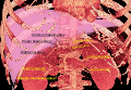 3D-rendered computed tomography, showing one renal artery (in whitish color) for each kidney, partially covered by the renal veins.
 -
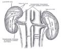
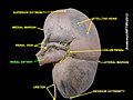
 Frontal section through the kidney
 -
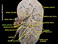
 Abdominal portion of the sympathetic trunk, with the celiac and hypogastric plexuses.
 -
 The posterior surfaces of the kidneys, showing areas of relation to the parietes.


-
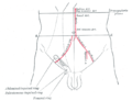 Front of abdomen, showing surface markings for arteries and inguinal canal.
 -
 Kidney
 -
 Renal artery
 -
 Renal artery
 -
 Renal artery
 -
 Renal artery
 -
 Renal artery


References
- ^ Renin-Dependent Hypertension Caused by Nonfocal Stenotic Aberrant Hypertension. 46(2):380-385, August 2005.Kem, David C.; Lyons, Daniel F.; Wenzl, James; Halverstadt, Donald; Yu, Xichun
- ^ Mathematical Modelling in Medicine. by Johnny T. Ottesen, Michael Danielsen - 2000 - Medical - 235 pages
- ^ http://emedicine.medscape.com/article/463015-overview Aytac SK, Yigit H, Sancak T, et al. Correlation between the diameter of the main renal artery and the presence of an accessory renal artery: sonographic and angiographic evaluation. J Ultrasound Med. May 2003;22(5):433-9; quiz 440-2.
External links
- MedlinePlus Image 9818
- Anatomy photo:40:11-0105 at the SUNY Downstate Medical Center - "Posterior Abdominal Wall: Branches of the Abdominal Aorta"
This cardiovascular system article is a stub. You can help Wikipedia by expanding it. |