
Warthin's tumor
| Warthin's tumor | |
|---|---|
| Specialty | Oncology |
- Not to be confused with Wharton's duct (submandibular duct)
Warthin's tumor or Warthin tumour, also known as papillary cystadenoma lymphomatosum or adenolymphoma, is a type of benign tumor of the salivary glands.
Etiology
Its etiology is unknown, but there is a strong association with cigarette smoking. Smokers are at 8 times greater risk of developing Warthin's tumor than the general population.[1]
Locations
The gland most likely affected is the parotid gland. Though much less likely to occur than pleomorphic adenoma, Warthin's tumor is the second most common benign parotid tumor.
Characteristic

Warthin's tumor primarily affects older individuals (age 60–70 years). There is a slight female predilection according to recent studies, but historically it has been associated with a strong male predilection. This change is possibly due to the tumor's association with cigarette smoking and the growing use of cigarettes by women. The tumor is slow growing, painless, and usually appears in the tail of the parotid gland near the angle of the mandible. In 5–14% of cases, Warthin's tumor is bilateral, but the two masses usually are at different times. Warthin's tumor is highly unlikely to become malignant.
Histology
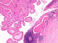
The appearance of this tumor under the microscope is unique. There are cystic spaces surrounded by two uniform rows of cells with centrally placed pyknotic nuclei. The cystic spaces have epithelium referred to as papillary infoldings that protrude into them. Additionally, the epithelium has lymphoid stroma with germinal center formation.
The differential diagnosis includes sebaceous lymphadenoma and oncocytoma.
Treatment
Most of these tumors are treated with surgical removal. It is non recurrent .
Additional images
-
 Histopathology of Warthin tumor in the parotid gland. H&E stain.
 -
 Histopathology of Warthin tumor in the parotid gland. Another view of a file "Warthin tumor (1).jpg". H&E stain.
 -
 Histopathology of Warthin tumor in the parotid gland. Higher magnification of a file "Warthin tumor (1).jpg". H&E stain.
 -
 Intermediate magnification micrograph of a Warthin tumor.
 -
 High magnification micrograph of a Warthin tumor showing the characteristic bilayered epithelium.
.jpg)
.jpg)
.jpg)


See also
References
- ^ Kumar V, Abbas AK, Fausto N (2005). Robbins and Cotran pathologic basis of disease, 7 Ed. St. Louis, MO: Elsevier Saunders. ISBN 0-7216-0187-1.
{{cite book}}: CS1 maint: multiple names: authors list (link)
Additional sources
- Kahn, Michael A. Basic Oral and Maxillofacial Pathology. Volume 1. 2001.
External links
This oncology article is a stub. You can help Wikipedia by expanding it. |