
Foveola: Difference between revisions
No edit summary |
|||
| Line 7: | Line 7: | ||
The centre of the foveola is sometimes referred to as the umbo. |
The centre of the foveola is sometimes referred to as the umbo. |
||
The anatomy of the foveola was recently reinvestigated.<ref name=pmid29576957>{{cite journal |doi=10.7717/peerj.4482 |pmid=29576957 |pmc=5853608 |title=The anatomy of the foveola reinvestigated |journal=Peerj |volume=6 |pages=e4482 |year=2018 |last1=Tschulakow |first1=Alexander V |last2=Oltrup |first2=Theo |last3=Bende |first3=Thomas |last4=Schmelzle |first4=Sebastian |last5=Schraermeyer |first5=Ulrich }} [[File:CC-BY icon.svg|50px]] Material was copied from this source, which is available under a [https://creativecommons.org/licenses/by/4.0/ Creative Commons Attribution 4.0 International License].</ref> |
|||
Serial semithin and ultrathin sections, and focused ion beam (FIB) tomography were prepared from 32 foveolae from monkeys (Macaca fascicularis) and humans. Serial sections and FIB analysis were then used to construct 3D models of central Müller and photoreceptor cells.<ref name=pmid29576957/> |
|||
It was discovered that in monkeys, outer segments of central foveolar cones are twice as long as those from parafoveal cones and do not run completely parallel to the incident light. Unique Müller cells are present in the central foveolae (area of 200 µm in diameter) of humans and monkeys.<ref name=pmid29576957/> |
|||
[[File:Macula.svg|thumb|Photograph of the retina of the human eye, with overlay diagrams showing the positions and sizes of the macula, fovea, and optic disc]] |
[[File:Macula.svg|thumb|Photograph of the retina of the human eye, with overlay diagrams showing the positions and sizes of the macula, fovea, and optic disc]] |
||
Revision as of 14:11, 13 August 2018
| Foveola | |
|---|---|
| Details | |
| Identifiers | |
| Latin | foveola |
| TA98 | A15.2.04.023 |
| TA2 | 6786 |
| FMA | 77666 |
| Anatomical terminology | |
The foveola is located within a region called the macula, a yellowish, cone photo receptor filled portion of the human retina. The foveola is approximately 0.35 mm in diameter and lies in the center of the fovea and contains only cone cells, and a cone-shaped zone of Müller cells.[1] In this region the cone receptors are found to be longer, slimmer and more densely packed than anywhere else in the retina, thus allowing that region to have the potential to have the highest visual acuity in the eye.
The centre of the foveola is sometimes referred to as the umbo.
The anatomy of the foveola was recently reinvestigated.[2]
Serial semithin and ultrathin sections, and focused ion beam (FIB) tomography were prepared from 32 foveolae from monkeys (Macaca fascicularis) and humans. Serial sections and FIB analysis were then used to construct 3D models of central Müller and photoreceptor cells.[2]
It was discovered that in monkeys, outer segments of central foveolar cones are twice as long as those from parafoveal cones and do not run completely parallel to the incident light. Unique Müller cells are present in the central foveolae (area of 200 µm in diameter) of humans and monkeys.[2]

Additional images
-
 Schematic diagram of the macula lutea of the retina, showing perifovea, parafovea, fovea, and clinical macula
 -
 An optical coherence tomography (OCT) scan of a macula at 800nm, with an axial resolution of 3µm
 -
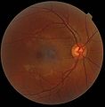 A fundus photograph showing the macula as a spot to the left. The optic disc is the area on the right where blood vessels converge. The grey, more diffuse spot in the centre is a shadow artifact.


Notes
- ^ Gass, J. Donald M (1999). "Müller Cell Cone, an Overlooked Part of the Anatomy of the Fovea Centralis". Archives of Ophthalmology. 117 (6): 821–3. doi:10.1001/archopht.117.6.821. PMID 10369597.
- ^ a b c Tschulakow, Alexander V; Oltrup, Theo; Bende, Thomas; Schmelzle, Sebastian; Schraermeyer, Ulrich (2018). "The anatomy of the foveola reinvestigated". Peerj. 6: e4482. doi:10.7717/peerj.4482. PMC 5853608. PMID 29576957.
{{cite journal}}: CS1 maint: unflagged free DOI (link) Material was copied from this source, which is available under a Creative Commons Attribution 4.0 International License.
| Fibrous tunic (outer) |
|  | |||||
|---|---|---|---|---|---|---|---|
| Uvea / vascular tunic (middle) |
| ||||||
| Retina (inner) |
| ||||||
| Anatomical regions of the eye |
| ||||||
| Other | |||||||
| Illusions |
| |
|---|---|---|
| Popular culture |
| |
| Related | ||
This article about the eye is a stub. You can help Wikipedia by expanding it. |