Breast: Difference between revisions
No edit summary |
|||
| Line 11: | Line 11: | ||
The breasts are modified sudoriferous (sweat) glands, producing [[milk]].<ref name="Tortora, Gerard J.">''Introduction to the Human Body, fifth ed.'' John Wiley & Sons, Inc.: New York, 2001. '''560'''</ref> |
The breasts are modified sudoriferous (sweat) glands, producing [[milk]].<ref name="Tortora, Gerard J.">''Introduction to the Human Body, fifth ed.'' John Wiley & Sons, Inc.: New York, 2001. '''560'''</ref> |
||
The breasts are covered by [[skin]]. Each breast has one [[nipple]] surrounded by the [[areola]]. The areola is colored from pink to dark brown and has several [[sebaceous gland]]s. The larger [[mammary gland]]s within the breast produce the milk. They are distributed throughout the breast, with two-thirds of the tissue found within 30 mm of the base of the nipple.<ref name="Ramsay">Anatomy of the lactating human breast redefined with ultrasound imaging, D.T. Ramsay et al., ''J. Anat.'' '''206''':525-534</ref> These are drained to the nipple by between 4 and 18 ''lactiferous ducts'', where each duct has its own opening. The network formed by these ducts is complex, like the tangled roots of a tree. It is not always arranged radially, and branches close to the nipple. The ducts near the nipple do not act as milk reservoirs. Ramsay '' et al.'' have shown that conventionally described ''lactiferous sinuses'' do not, in fact, exist. |
LOLOLOLOLZ!!!!The breasts are covered by [[skin]]. Each breast has one [[nipple]] surrounded by the [[areola]]. The areola is colored from pink to dark brown and has several [[sebaceous gland]]s. The larger [[mammary gland]]s within the breast produce the milk. They are distributed throughout the breast, with two-thirds of the tissue found within 30 mm of the base of the nipple.<ref name="Ramsay">Anatomy of the lactating human breast redefined with ultrasound imaging, D.T. Ramsay et al., ''J. Anat.'' '''206''':525-534</ref> These are drained to the nipple by between 4 and 18 ''lactiferous ducts'', where each duct has its own opening. The network formed by these ducts is complex, like the tangled roots of a tree. It is not always arranged radially, and branches close to the nipple. The ducts near the nipple do not act as milk reservoirs. Ramsay '' et al.'' have shown that conventionally described ''lactiferous sinuses'' do not, in fact, exist. |
||
The remainder of the breast is composed of [[connective tissue]] ([[collagen]] and [[elastin]]), [[adipose tissue]] (fat), and [[Cooper's ligaments|Cooper’s ligaments]]. The ratio of glands to adipose tissues rises from 1:1 in nonlactating women to 2:1 in lactating women.<ref name="Ramsay"/> |
The remainder of the breast is composed of [[connective tissue]] ([[collagen]] and [[elastin]]), [[adipose tissue]] (fat), and [[Cooper's ligaments|Cooper’s ligaments]]. The ratio of glands to adipose tissues rises from 1:1 in nonlactating women to 2:1 in lactating women.<ref name="Ramsay"/> |
||
Revision as of 18:57, 16 February 2007

The term breast refers to the upper ventral region of an animal’s torso, particularly that of mammals, including human beings. The breasts of a female mammal’s body contain the mammary glands, which secrete milk used to feed infants. This article focuses on human female breasts, but male humans also have breasts which are usually less prominent, but structurally identical and homologous to the female, as they develop embryologically from the same tissues. In some situations male breast development does occur, a condition called gynecomastia.
Anatomy

The breasts are modified sudoriferous (sweat) glands, producing milk.[1]
LOLOLOLOLZ!!!!The breasts are covered by skin. Each breast has one nipple surrounded by the areola. The areola is colored from pink to dark brown and has several sebaceous glands. The larger mammary glands within the breast produce the milk. They are distributed throughout the breast, with two-thirds of the tissue found within 30 mm of the base of the nipple.[2] These are drained to the nipple by between 4 and 18 lactiferous ducts, where each duct has its own opening. The network formed by these ducts is complex, like the tangled roots of a tree. It is not always arranged radially, and branches close to the nipple. The ducts near the nipple do not act as milk reservoirs. Ramsay et al. have shown that conventionally described lactiferous sinuses do not, in fact, exist.
The remainder of the breast is composed of connective tissue (collagen and elastin), adipose tissue (fat), and Cooper’s ligaments. The ratio of glands to adipose tissues rises from 1:1 in nonlactating women to 2:1 in lactating women.[2]
The breasts sit over the pectoralis major muscle and usually extend from the level of the 2nd rib to the level of the 6th rib anteriorly. The superior lateral quadrant of the breast extends diagonally upwards towards the axillae and is known as the tail of Spense. A thin layer of mammary tissue extends from the clavicle above to the seventh or eighth ribs below and from the midline to the edge of the latissimus dorsi posteriorly.
The arterial blood supply to the breasts is derived from the internal thoracic artery (formerly called the internal mammary artery), lateral thoracic artery, thoracoacromial artery, and posterior intercostal arteries. The venous drainage of the breast is mainly to the axillary vein, but there is some drainage to the internal thoracic vein and the intercostal veins. Both sexes have a large concentration of blood vessels and nerves in their nipples.
The breast is innervated by the anterior and lateral cutaneous branches of the fourth through sixth intercostal nerves. The nipple is supplied by the T4 dermatome.
The primary anatomical support for the breasts is thought to be provided by the Cooper’s ligaments, with additional support from the skin covering the breasts themselves, and it is this support which determines the shape of the breasts. The external shape or size of the breast is not predictive of its internal anatomy nor of its lactation potential. In a small fraction of women, the frontal milk sinuses (ampulla) in the breasts are not flush with the surrounding breast tissue, which causes the sinus area to visibly bulge outward.
Lymphatic drainage
About 75 percent of lymph from the breast travels to the ipsilateral axillary lymph nodes. The rest travels to parasternal nodes, to the other breast, or abdominal lymph nodes. The axillary nodes include the pectoral, subscapular, and humeral groups of lymph nodes. These drain to the central axillary lymph nodes, then to the apical axillary lymph nodes. The lymphatic drainage of the breasts is particularly relevant to oncology, since breast cancer is a common cancer and cancer cells can break away from a tumour and spread to other parts of the body through the lymph system by metastasis.
Function
Breastfeeding and pregnancy

The function of mammary glands in female breasts is to nurture young by producing milk, which is secreted by the nipples during lactation. While the mammary glands that produce milk are present in the male, they normally remain undeveloped. The orb-like shape of breasts may help limit heat loss, as a fairly high temperature is required for the production of milk. During lactation, the energy consumption of the breast exceeds that of the brain,[citation needed] otherwise by far the body’s most energy-hungry organ. Alternatively, the human breast evolved in order to prevent infants from suffocating while feeding. Since human infants do not have a protruding jaw like human evolutionary ancestors and other primates, the infant’s nose might be blocked by a flat female chest while feeding. According to this theory, as the human jaw receded, the breasts became larger to compensate.[4]
Milk production can also occur in both men and women as an adverse effect of some medicinal drugs (such as some antipsychotic medication), extreme physical stress or in endocrine disorders. Newborn babies are often capable of lactation because they receive some amount of prolactin and oxytocin (milk hormones) from their connection to the mother.
Other suggested functions
Zoologists point out that no female mammal other than the human has breasts of comparable size when not lactating and that humans are the only primate that have permanently swollen breasts. This suggests that the external form of the breasts is connected to factors other than lactation alone.
One theory is based around the fact that, unlike nearly all other primates, human females do not display clear, physical signs of ovulation. This could have plausibly resulted in human males evolving to respond to more subtle signs of ovulation. During ovulation, the increased estrogen present in the female body results in a slight swelling of the breasts, which then males could have evolved to find attractive. In response, there would be evolutionary pressures that would favor females with more swollen breasts who would, in a manner of speaking, appear to males to be the most likely to be ovulating.
Some zoologists (notably Desmond Morris) believe that the shape of female breasts evolved as a frontal counterpart to that of the buttocks, the reason being that whilst other primates mate in the rear-entry position, humans are more likely to successfully copulate mating face on. A secondary sexual characteristic on a woman’s chest would have encouraged this in more primitive incarnations of the human race, and a face on encounter may have helped found a relationship between partners beyond merely a sexual one.[5]
Size and shape
Shape and support
Aside from size variations, there is naturally large variety in the shape of breasts. As with all body parts, the shape of the breast is determined primarily by the differential rate of growth in different areas, and the direction of said growth. Additionally, the shape of a woman’s breast is in large part dependent on their support, which primarily comes from the skin and the ligaments of the breasts themselves, and the underlying chest on which they rest. The breast is attached at its base to the chest wall by the deep fascia over the pectoral muscles. On its upper surface it is suspended by the covering skin where it continues on to the upper chest wall.
In discussing the support of breasts, it is helpful to draw a distinction between breasts which extend below (the inframammary line) to rest on the chest below, and those which do not. Breasts which do not extend below the inframammary line at all form a rounded dome shape protruding almost horizontally from the chest wall. All breasts are like this in early stages of development, and such a shape is common in younger women and girls. This protruding or “high” breast is anchored to the chest at its base, and the weight is distributed evenly over the area of the base of the approximately cone-shaped breasts.

In the “low” breast, a proportion of the breasts’ weight is actually supported by the chest against which the lower breast surface comes to rest, as well as the deep anchorage at the base. The weight is thus distributed over a larger area. This has the effect of reducing the strain. In both males and females, the thoracic cavity slopes progressively outwards from the thoracic inlet (at the top of the breastbone) above to the lowest ribs which mark its lower boundary, allowing it to support the breasts.
The inframammary fold (IMF) is an anatomic structure created by adherence between elements in the skin and underlying connective tissue[6] and represents the inferior extent of breast anatomy. The relationship of the nipple positon to this IMF is described as ptosis. Due to breast weight and relaxation of support structures, the NAC and breast tissue may eventually hang below the IMF, and in some cases the breasts may extend as far as, or even beyond, the navel. The length from the nipple to the sternal notch (central, upper border) in the youthful breast averages 21 cm and is a common anthropometric figure used to assess both breast symmetry and ptosis. Lengthening of both this measurement and the distance between the NAC and IMF are both characteristic of advancing grades of ptosis. Some teenagers may develop breasts whose skin comes into contact with the chest below the inframammary fold at an early age, and some women may never develop such breasts. Both situations are perfectly normal.
The end of the breast, which includes the nipple, may either be flat (a 180 degree angle) or angled (angles lower than 180 degrees). Breast ends are rarely angled sharper than 60 degrees. Angling of the end of the breast is caused in part by the ligaments that suspend it, such that the breast ends often have a more obtuse angle when a woman is lying on her back. Breasts exist in a range of ratios between length and base diameter, usually ranging from 1/2 to 1.
Additional, external support
Since the breasts are flexible, the shape of breasts may be strongly affected by clothing, and foundation garments in particular. A bra may be worn to give additional support and to alter the shape of the breasts. There is some debate over whether such support is desirable. A long term clinical study showed that women with large breasts can suffer shoulder pain as a result of bra straps,[7] although a well fitting bra should support most of the breasts’ weight with proper sized cups and back band rather than on the shoulders. (See Myalgia (brassiere).)
Changes

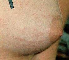
As breasts are mostly composed of adipose tissue, their size can change over time. This occurs for a number of reasons, for example if the woman gains or loses weight. Any rapid increase in size of the breasts, during puberty, weight gain or pregnancy, can result in the appearance of stretchmarks on the skin.
It is also typical for a number of changes to occur during pregnancy: the breasts generally become larger and firmer, mainly due to hypertrophy of the mammary gland in response to the hormone prolactin. The size of the nipples may increase noticeably and their pigmentation may become darker. These changes may continue during breastfeeding. The breasts generally revert to approximately their previous size after pregnancy, although there may be some increased sagging and stretchmarks.
The size of a woman’s breasts usually also fluctuates during the menstrual cycle, particularly with premenstrual water retention. An increase in breast size is also a common side effect of use of the combined oral contraceptive pill.
The breasts naturally sag through aging, as the ligaments become elongated. This process may be accelerated by high impact exercises, and a brassiere may reduce this effect by providing external support, although the health benefits of wearing of a brassiere are not universally accepted.
Some women undergo breast reconstruction after mastectomy for breast cancer, a result of the high value placed on symmetry of the human form in those cultures, and because women often identify their femininity and sense of self with their breasts.
Plastic surgery
Plastic surgical procedures of the breast include those for both cosmetic and reconstructive surgery indications. After mastectomy some women choose to have their breasts reconstructed, either with breast implants or autologous tissue transfer, using fat and tissues from the abdomen (TRAM flap) or back (latissiumus muscle flap). Breast reduction surgery is a common procedure which involves resecting excess breast tissue and skin with repositioning of the nipple-areolar complex (NAC). Cosmetic procedures include breast lifts (mastopexy), breast augmentation with implants, and procedures that combine both elements. Implants containing either silicone gel or saline are available for augmentation and reconstructive surgeries.
Any surgery of the breast carries with it the potential for interfering with future breastfeeding [8][9][10], causing alterations in NAC sensation, and possible difficulty in interpreting mammography (xrays of the breast). A number of studies have demonstrated a similar ability to breastfeed when breast reduction patients are compared to control groups where the surgery was performed using a modern pedicle surgical technique. [11][12][13] [14]Plastic Surgery organizations have generally discouraged elective cosmetic breast surgery in teens as the volume of their breast tissue may continue to grow significantly as they mature and over concerns of understanding long-term risks and benefits of the procedure. [2]
Development
The development of a woman’s breasts during puberty is triggered by sex hormones, chiefly estrogen. This hormone has been demonstrated to cause the development of woman-like, enlarged breasts in men, a condition called gynecomastia, and is sometimes used deliberately for this effect in male-to-female hormone replacement therapy.
In most cases, the breasts do fold down over the chest wall during development, as shown in this diagram.[15] It is typical for a woman’s breasts to be unequal in size particularly while the breasts are developing during puberty. Statistically it is slightly more common for the left breast to be the larger.[16] In rare cases, the breasts may be significantly different in size, or one breast may fail to develop entirely.
A vast number of medical conditions are known to cause abnormal development of the breasts during puberty. Virginal breast hypertrophy is a condition which involves excessive growth of the breasts during puberty, and in some cases the continued growth beyond the usual pubescent age. Breast hypoplasia is a condition where one or both breasts fail to develop during puberty.
In Cameroon, some girls are subjected to breast ironing to stunt breast growth in order to make them less sexually attractive and thus become less likely to become a victim of rape.
Cultural status

Historically, breasts have been regarded as fertility symbols, because they are the source of life-giving milk. Certain prehistoric female statuettes—so-called Venus figurines—often emphasised the breasts, as in the example of the Venus of Willendorf. In historic times, goddesses such as Ishtar were shown with many breasts, alluding to their role as goddesses of childbirth and mothering.
Breasts are secondary sex characteristics and sexually sensitive. Bare female breasts can elicit heightened sexual desires from men and women. Since they are associated with sex, in many cultures bare breasts are indecent, and they are not commonly displayed in public, in contrast to male chests. Other cultures view the baring of breasts as acceptable, and in some countries women have never been forbidden to bare their chests. Opinion on the exposure of breasts is often dependent on the place and context, and in some Western societies exposure of breasts on a beach may be acceptable, although in town centres, for example, it is usually indecent. In some areas, the prohibition against the display of a woman’s breasts generally only restricts exposure of the nipples.
When breastfeeding a baby in public, legal and social rules regarding indecent exposure and dress code, as well as inhibitions of the woman, tend to be relaxed. Numerous laws around the world have made public breastfeeding legal and disallow companies from prohibiting it in the workplace. Yet the public reaction at the sight of breastfeeding can make the situation uncomfortable for those involved.
Women in some areas and cultures are approaching the issue of breast exposure as one of sexual equality, since men (and pre-pubescent children) may bare their chests, but women and teenage girls are forbidden. In the United States, the topfree equality movement seeks to redress this imbalance. This movement won a decision in 1992 in the New York State Court of Appeals—“People v. Santorelli”, where the court ruled that the state’s indecent exposure laws do not ban women from being barebreasted. A similar movement succeeded in most parts of Canada in the 1990s. In Australia and much of Europe it is acceptable for women and teenage girls to sunbathe topless on some public beaches and swimming pools, but these are generally the only public areas where exposing breasts is acceptable.
Some religions require that women always keep their breasts covered. For example, Islam forbids public exposure of the female breasts.[17]
In addition to the above references, see also modesty, nudism and exhibitionism.
In some paintings women are shown with their breasts in their hands or on a platter, signifying that they died as a martyr by having their breasts severed. One example of this is Saint Agatha.
Disorders
Infections and inflammations
These may be caused among others by trauma, secretory stasis/milk engorgment, hormonal stimulation, infections or autoimmune reactions. Repeated occurrence unrelated to lactation requires endocrinological examination.
- Mastitis
- bacterial mastitis
- mastitis from milk engorgement or secretory stasis
- mastitis of mumps
- chronic intramammary abscess
- chronic subareolar abscess
- tuberculosis of the breast
- syphilis of the breast
- retromammary abscess
- actinomycosis of the breast
- Mondor’s disease
- duct ectasia syndrome
- breast engorgement
Benign diseases
- Congenital disorders
- inverted nipple
- supernumerary nipples/supernumerary breasts (polymazia / polymastia)/duplicated nipples
- Aberrations of normal development and involution
- cyclical nodularity
- cysts
- fibroadenoma - benign tumor
- nipple discharge, galactorhea - not allways a disease
- mammary fistula
- Fibrocystic disease/Fibrocystic changes
- cysts
- epithelial hyperplasia
- epithelial metaplasia
- papillomas
- adenosis
- Pregnancy-related
Malignant diseases
- Breast cancer (mammary carcinoma)
- Carcinoma in situ
- Paget’s disease of the nipple, also known as Paget’s disease of the breast
References
- ^ Introduction to the Human Body, fifth ed. John Wiley & Sons, Inc.: New York, 2001. 560
- ^ a b Anatomy of the lactating human breast redefined with ultrasound imaging, D.T. Ramsay et al., J. Anat. 206:525-534
- ^ A Woman's Body: Breasts are Not Just for Filling Sweaters Article
- ^ Bentley, Gillian R. (2001). "The Evolution of the Human Breast". American Journal of Physical Anthropology. 32 (38).
- ^ Morris, Desmond (1967). The Naked Ape: a zoologist's study of the human animal. Canada: Bantam Books. pp. pp. 64-68. N3924.
{{cite book}}:|pages=has extra text (help) - ^ Boutros S, Kattash M, Wienfeld A, Yuksel E, Baer S, Shenaq S. The intradermal anatomy of the inframammary fold. Plast Reconstr Surg. 1998 Sep;102(4):1030-3. PMID 9734420
- ^ Ryan, EL, Pectoral girdle myalgia in women: a 5-year study in a clinical setting. Clin J Pain. 2000 Dec;16(4):298-303
- ^ Neifert, M (1990). "The influence of breast surgery, breast appearance and pregnancy-induced changes on lactation sufficiency as measured by infant weight gain". Birth. 17 (1): pp. 31-38. PMID 2288566.
{{cite journal}}:|access-date=requires|url=(help);|pages=has extra text (help); Unknown parameter|coauthors=ignored (|author=suggested) (help) - ^ "FAQ on Previous Breast Surgery and Breastfeeding". La Leche League International. 2006-08-29. Retrieved 2007-02-11.
- ^ West, Diana. "Breastfeeding After Breast Surgery". Australian Breastfeeding Association. Retrieved 2007-02-11.
- ^ Cruz-Korchin, N (2004-09-15). "Breast-feeding after vertical mammaplasty with medial pedicle". Plast Reconstr Surg. 15 (114): pp. 890-894. PMID 15468394.
{{cite journal}}:|access-date=requires|url=(help);|pages=has extra text (help); Unknown parameter|coauthors=ignored (|author=suggested) (help) - ^ Brzozowski, D (February 2000). "Breast-feeding after inferior pedicle reduction mammaplasty". Plast Reconstr Surg. 105 (2): pp. 530-534. PMID 10697157.
{{cite journal}}:|access-date=requires|url=(help);|pages=has extra text (help); Unknown parameter|coauthors=ignored (|author=suggested) (help) - ^ Witte, PM (2004-06-26). "Successful breastfeeding after reduction mammaplasty". Ned Tijdschr Geneeskd. 148 (26): pp. 1291-1293. PMID 15279213.
{{cite journal}}:|access-date=requires|url=(help);|pages=has extra text (help); Unknown parameter|coauthors=ignored (|author=suggested) (help) - ^ Kakagia, D (2005-10). "Breastfeeding after reduction mammaplasty: a comparison of 3 techniques". Ann Plast Surg. 55 (4): pp. 343-345. PMID 16186694.
{{cite journal}}:|access-date=requires|url=(help);|pages=has extra text (help); Check date values in:|date=(help); Unknown parameter|coauthors=ignored (|author=suggested) (help) - ^ A.R. Greenbaum, T. Heslop, J. Morris and K.W. Dunn, An investigation of the suitability of bra fit in women referred for reduction mammaplasty, Br J Plast Surg 56 (2003) (3), pp. 230–236
- ^ C.W. Loughry; et al. (1989). "Breast volume measurement of 598 women using biostereometric analysis". Annals of Plastic Surgery. 22 (5): 380–385.
{{cite journal}}: Explicit use of et al. in:|author=(help) - ^ “They shall cover their chests” or “they should draw their khimar (veils) over their bosoms”, depending on the translation, Quran (24:31)[1]
See also
- Brassiere
- Breastfeeding
- Breast fetishism
- Breast cancer
- Breast implant
- Breast reconstruction
- Breast self-examination
- Gynecomastia
- Intimate part
- Mammary intercourse
- Male lactation
- Puberty
- Tanner stage
- Topfree equality
- Toplessness
- Women
