Anterior ciliary arteries
| Anterior ciliary arteries | |
|---|---|
 | |
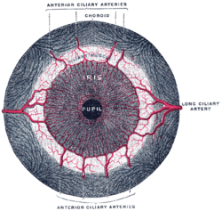
 Iris, front view. | |
| Details | |
| Source | Ophthalmic artery |
| Vein | Anterior ciliary veins |
| Supplies | Conjunctiva, sclera and rectus muscles |
| Identifiers | |
| Latin | Arteriae ciliares anteriores |
| TA98 | A12.2.06.034 |
| TA2 | 4485 |
| FMA | 70782 |
| Anatomical terminology | |
The anterior ciliary arteries are seven small arteries in each eye-socket that supply the conjunctiva, sclera and the rectus muscles. They are derived from the muscular branches of the ophthalmic artery.
Course
The anterior ciliary arteries are branches of the ophthalmic artery and run to the front of the eyeball in company with the extraocular muscles. They form a vascular zone beneath the conjunctiva, and then pierce the sclera a short distance from the cornea and end in the circulus arteriosus major. Three of the four rectus muscles; the superior, inferior and medial, are supplied by two ciliary arteries each, while the lateral rectus only receives one branch.
References
![]() This article incorporates text in the public domain from page 571 of the 20th edition of Gray's Anatomy (1918)
This article incorporates text in the public domain from page 571 of the 20th edition of Gray's Anatomy (1918)
External links
