
Lateral sulcus
| Lateral sulcus | |
|---|---|
 Lateral sulcus | |
| Details | |
| Identifiers | |
| Latin | fissura lateralis cerebri, sulcus lateralis cerebri |
| NeuroNames | 49 |
| NeuroLex ID | birnlex_1487 |
| TA98 | A14.1.06.006 A14.1.09.104 |
| TA2 | 5436, 5877 |
| FMA | 77801 |
| Anatomical terms of neuroanatomy | |
The lateral sulcus (also called Sylvian fissure or lateral fissure) is one of the most prominent structures of the human brain.
Anatomy
The lateral sulcus divides both the frontal lobe and parietal lobe above from the temporal lobe below. It's in both hemispheres of the brain but is longer in the left hemisphere in most people. The lateral sulcus is one of the earliest-developing sulci of the human brain. It first appears around the fourteenth gestational week.[1]
The lateral sulcus has a number of side branches. Two of the most prominent and most regularly found are the ascending (also called vertical) ramus and the horizontal ramus of the lateral fissure, which subdivide the inferior frontal gyrus. The lateral sulcus also contains the transverse temporal gyri, which are part of the primary and below the surface auditory cortex.
Partly due to a phenomenon called Yakovlevian torque, the lateral sulcus is often longer and less curved on the left hemisphere than on the right.
It is also located near Sylvian Point.
The area lying around the Sylvian fissure is often referred to as the perisylvian cortex.[2]
The human secondary somatosensory cortex (S2, SII) is a functionally-defined region of cortex in the parietal operculum on the ceiling of the lateral sulcus.
Discovery

The cerebral cortex was not depicted in a realistic manner until the 17th century with the Sylvian fissure being first accurately painted by Girolamo Fabrici d'Acquapendente in 1600 to provide plates for his Tabulae Pictae.[3][4][5]

Its first description is traditionally taken to be in 1641 by Caspar Bartholin who attributed its discovery to Franciscus Sylvius (1614–1672), professor of medicine at Leiden University his book Casp. Bartolini Institutiones Anatomicae where it is noted that "F.S. [F.S. probably refers to Franciscus Sylvius] If you examine the indentations which are represented in Figure 5 quite attentively, you will notice that they are very deep and that the brain is divided from one side to the other by the “anfractuosa fissura,” which starts in the front part near the ocular roots, and from there moves backwards above the base of the spinal cord, following the temporal bones, and it divides the upper part of the brain from the lower."[3]
It has been suggested that since Caspar Bartholin died in 1629 and Franciscus Sylvius only started medicine in 1632 that these words are either by his son Thomas Bartholin or Franciscus Sylvius. In 1663 in his Disputationem Medicarum, Franciscus Sylvius described the lateral fissure: "Particularly noticeable is the deep fissure or hiatus which begins at the roots of the eyes (oculorum radices) . . . it runs posteriorly above the temples as far as the roots of the brain stem (medulla radices). . . . It divides the cerebrum into an upper, larger part and a lower, smaller part".[3]
Additional images
-
 Base of brain (lateral fissure visible at top left)
 -
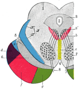 Coronal section through mid-brain
 -
 Posterior and inferior cornua of left lateral ventricle exposed from the side
 -
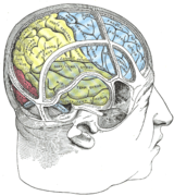 Drawing to illustrate the relations of the brain to the skull
 -
 Relations of the brain and middle meningeal artery to the surface of the skull
 -
 Human brain frontal (coronal) section: #24 is lateral sulcus.
 -
 Lateral sulcus shown in red (animation)
 -
 Cerebrum. Lateral view. Deep dissection.
 -
 Cerebrum. Lateral view. Deep dissection.
 -
 Cerebrum. Lateral view. Deep dissection.
 -
 Cerebrum. Optic and olfactory nerves. Inferior view. Deep dissection.
 -
 Cerebrum.Inferior view. Deep dissection.


_section_description_2.JPG)


References
- ^ Jee G. Chi, Elizabeth C. Dooling, Floyd H. Gilles (January 1977). "Gyral development of the human brain". Annals of Neurology. 1 (1): 86–93. doi:10.1002/ana.410010109. PMID 560818.
{{cite journal}}: CS1 maint: multiple names: authors list (link) - ^ Courten Norbury: Understanding Developmental Language Disorders: From Theory to Practice 2008, p. 63
- ^ a b c Collice, M; Collice, R; Riva, A. (2008). "Who discovered the sylvian fissure?". Neurosurgery. 63 (4): 623–8. doi:10.1227/01.NEU.0000327693.86093.3F. PMID 18981875.
- ^ Zanchin, G.; De Caro, R. (Oct 2006). "The nervous system in colours: the tabulae pictae of G.F. d'Acquapendente (ca. 1533-1619)". J Headache Pain. 7 (5): 360–6. doi:10.1007/s10194-006-0340-0. PMC 3468184. PMID 17058037.
- ^ Riva, A. (Sep 2007). "G.F. d'Acquapendente tabulae pictae on the nervous system". J Headache Pain. 8 (4): 253–4, author reply 255-6. doi:10.1007/s10194-007-0408-5. PMC 3451669. PMID 17906833.