
Cyanobacteria: Difference between revisions
Epipelagic (talk | contribs) |
Epipelagic (talk | contribs) add stuff on motility, including copy from Synechococcus – see that page's history for attribution |
||
| Line 178: | Line 178: | ||
===Cellular death=== |
===Cellular death=== |
||
One of the most critical processes determining cyanobacterial eco-physiology is [[cellular death]]. Evidence supports the existence of controlled cellular demise in cyanobacteria, and various forms of cell death have been described as a response to biotic and abiotic stresses. However, cell death research in this phylogenetic group is a relatively young field and understanding of the underlying mechanisms and molecular machinery underpinning this fundamental process remains largely elusive.<ref name=Aguilera2021 /> However, reports on cell death of marine and freshwater cyanobacteria indicate that this process has major implications for the ecology of microbial communities/<ref name="Agustí2004">Agustí, S. (2004). [https://digital.csic.es/handle/10261/86957 "Viability and niche segregation of ''Prochlorococcus'' and ''Synechococcus'' cells across the Central Atlantic Ocean."] Accessed: 30 July 2021).</ref><ref name="Agustí2006">{{cite journal |doi = 10.1111/j.1365-2427.2006.01584.x|title = Cell death in lake phytoplankton communities|year = 2006|last1 = Agusti|first1 = Susana|last2 = Alou|first2 = EVA|last3 = Hoyer|first3 = Mark V.|last4 = Frazer|first4 = Thomas K.|last5 = Canfield|first5 = Daniel E.|journal = Freshwater Biology|volume = 51|issue = 8|pages = 1496–1506}}</ref><ref name=Franklin2006>{{cite journal |doi = 10.1080/09670260500505433|title = What is the role and nature of programmed cell death in phytoplankton ecology?|year = 2006|last1 = Franklin|first1 = Daniel J.|last2 = Brussaard|first2 = Corina P.D.|last3 = Berges|first3 = John A.|journal = European Journal of Phycology|volume = 41|pages = 1–14|s2cid = 53599616}}</ref><ref name=Sigee2007>{{cite journal |doi = 10.2216/06-69.1|title = Patterns of cell death in freshwater colonial cyanobacteria during the late summer bloom|year = 2007|last1 = Sigee|first1 = D. C.|last2 = Selwyn|first2 = A.|last3 = Gallois|first3 = P.|last4 = Dean|first4 = A. P.|journal = Phycologia|volume = 46|issue = 3|pages = 284–292|s2cid = 86268392}}</ref> Different forms of cell demise have been observed in cyanobacteria under several stressful conditions,<ref name=BermanFrank2004>{{cite journal |doi = 10.4319/lo.2004.49.4.0997|title = The demise of the marine cyanobacterium, Trichodesmium SPP., via an autocatalyzed cell death pathway|year = 2004|last1 = Berman-Frank|first1 = Ilana|last2 = Bidle|first2 = Kay D.|last3 = Haramaty|first3 = Liti|last4 = Falkowski|first4 = Paul G.|journal = Limnology and Oceanography|volume = 49|issue = 4|pages = 997–1005|bibcode = 2004LimOc..49..997B}}</ref><ref name=Hu2019>{{cite journal |doi = 10.3390/toxins11120706|doi-access = free|title = Programmed Cell Death-Like and Accompanying Release of Microcystin in Freshwater Bloom-Forming Cyanobacterium Microcystis: From Identification to Ecological Relevance|year = 2019|last1 = Hu|first1 = Chenlin|last2 = Rzymski|first2 = Piotr|journal = Toxins|volume = 11|issue = 12|page = 706|pmid = 31817272|pmc = 6950475}}</ref> and cell death has been suggested to play a key role in developmental processes, such as akinete and heterocyst differentiation.<ref>{{cite journal |doi = 10.1023/A:1013840025518|year = 2001|last1 = Meeks|first1 = John C.|last2 = Elhai|first2 = Jeff|last3 = Thiel|first3 = Teresa|last4 = Potts|first4 = Malcolm|last5 = Larimer|first5 = Frank|last6 = Lamerdin|first6 = Jane|last7 = Predki|first7 = Paul|last8 = Atlas|first8 = Ronald|title = An overview of the genome of Nostoc punctiforme, a multicellular, symbiotic cyanobacterium|journal = Photosynthesis Research|volume = 70|issue = 1|pages = 85–106|pmid = 16228364|s2cid = 8752382}}</ref><ref name=Claessen2014 /><ref name=Aguilera2021 /> |
One of the most critical processes determining cyanobacterial eco-physiology is [[cellular death]]. Evidence supports the existence of controlled cellular demise in cyanobacteria, and various forms of cell death have been described as a response to biotic and abiotic stresses. However, cell death research in this phylogenetic group is a relatively young field and understanding of the underlying mechanisms and molecular machinery underpinning this fundamental process remains largely elusive.<ref name=Aguilera2021 /> However, reports on cell death of marine and freshwater cyanobacteria indicate that this process has major implications for the ecology of microbial communities/<ref name="Agustí2004">Agustí, S. (2004). [https://digital.csic.es/handle/10261/86957 "Viability and niche segregation of ''Prochlorococcus'' and ''Synechococcus'' cells across the Central Atlantic Ocean."] Accessed: 30 July 2021).</ref><ref name="Agustí2006">{{cite journal |doi = 10.1111/j.1365-2427.2006.01584.x|title = Cell death in lake phytoplankton communities|year = 2006|last1 = Agusti|first1 = Susana|last2 = Alou|first2 = EVA|last3 = Hoyer|first3 = Mark V.|last4 = Frazer|first4 = Thomas K.|last5 = Canfield|first5 = Daniel E.|journal = Freshwater Biology|volume = 51|issue = 8|pages = 1496–1506}}</ref><ref name=Franklin2006>{{cite journal |doi = 10.1080/09670260500505433|title = What is the role and nature of programmed cell death in phytoplankton ecology?|year = 2006|last1 = Franklin|first1 = Daniel J.|last2 = Brussaard|first2 = Corina P.D.|last3 = Berges|first3 = John A.|journal = European Journal of Phycology|volume = 41|pages = 1–14|s2cid = 53599616}}</ref><ref name=Sigee2007>{{cite journal |doi = 10.2216/06-69.1|title = Patterns of cell death in freshwater colonial cyanobacteria during the late summer bloom|year = 2007|last1 = Sigee|first1 = D. C.|last2 = Selwyn|first2 = A.|last3 = Gallois|first3 = P.|last4 = Dean|first4 = A. P.|journal = Phycologia|volume = 46|issue = 3|pages = 284–292|s2cid = 86268392}}</ref> Different forms of cell demise have been observed in cyanobacteria under several stressful conditions,<ref name=BermanFrank2004>{{cite journal |doi = 10.4319/lo.2004.49.4.0997|title = The demise of the marine cyanobacterium, Trichodesmium SPP., via an autocatalyzed cell death pathway|year = 2004|last1 = Berman-Frank|first1 = Ilana|last2 = Bidle|first2 = Kay D.|last3 = Haramaty|first3 = Liti|last4 = Falkowski|first4 = Paul G.|journal = Limnology and Oceanography|volume = 49|issue = 4|pages = 997–1005|bibcode = 2004LimOc..49..997B}}</ref><ref name=Hu2019>{{cite journal |doi = 10.3390/toxins11120706|doi-access = free|title = Programmed Cell Death-Like and Accompanying Release of Microcystin in Freshwater Bloom-Forming Cyanobacterium Microcystis: From Identification to Ecological Relevance|year = 2019|last1 = Hu|first1 = Chenlin|last2 = Rzymski|first2 = Piotr|journal = Toxins|volume = 11|issue = 12|page = 706|pmid = 31817272|pmc = 6950475}}</ref> and cell death has been suggested to play a key role in developmental processes, such as akinete and heterocyst differentiation.<ref>{{cite journal |doi = 10.1023/A:1013840025518|year = 2001|last1 = Meeks|first1 = John C.|last2 = Elhai|first2 = Jeff|last3 = Thiel|first3 = Teresa|last4 = Potts|first4 = Malcolm|last5 = Larimer|first5 = Frank|last6 = Lamerdin|first6 = Jane|last7 = Predki|first7 = Paul|last8 = Atlas|first8 = Ronald|title = An overview of the genome of Nostoc punctiforme, a multicellular, symbiotic cyanobacterium|journal = Photosynthesis Research|volume = 70|issue = 1|pages = 85–106|pmid = 16228364|s2cid = 8752382}}</ref><ref name=Claessen2014 /><ref name=Aguilera2021 /> |
||
{{clear}} |
|||
==Motility== |
|||
[[File:Cyanobacteriaunicellularandcolonial020 Synechococcus.jpg|thumb|upright=1| ''[[Synechococcus]]'' uses a gliding technique to move at 25 μm/s. Scale bar: about 10 µm.]] |
|||
It has long been known that [[filamentous cyanobacteria]] perform surface motions, and that these movements result from [[type IV pili]].<ref>{{cite journal |doi = 10.1128/JB.01927-06|title = Molecular Analysis of Genes in Nostoc punctiforme Involved in Pilus Biogenesis and Plant Infection|year = 2007|last1 = Duggan|first1 = Paula S.|last2 = Gottardello|first2 = Priscila|last3 = Adams|first3 = David G.|journal = Journal of Bacteriology|volume = 189|issue = 12|pages = 4547–4551|pmid = 17416648|pmc = 1913353}}</ref><ref>{{cite journal |doi = 10.1111/mmi.13205|title = Evidence that a modified type IV pilus-like system powers gliding motility and polysaccharide secretion in filamentous cyanobacteria|year = 2015|last1 = Khayatan|first1 = Behzad|last2 = Meeks|first2 = John C.|last3 = Risser|first3 = Douglas D.|journal = Molecular Microbiology|volume = 98|issue = 6|pages = 1021–1036|pmid = 26331359|s2cid = 8749419}}</ref><ref>{{cite journal |doi = 10.1111/mmi.13242|title = Motility in cyanobacteria: Polysaccharide tracks and Type IV pilus motors|year = 2015|last1 = Wilde|first1 = Annegret|last2 = Mullineaux|first2 = Conrad W.|journal = Molecular Microbiology|volume = 98|issue = 6|pages = 998–1001|pmid = 26447922|s2cid = 22585994}}</ref> Additionally, ''[[Synechococcus]]'', a marine cyanobacteria, is known to swim at a speed of 25 μm/s by a mechanism different to that of bacterial flagella.<ref>{{cite journal |doi = 10.1126/science.230.4721.74|title = A Cyanobacterium Capable of Swimming Motility|year = 1985|last1 = Waterbury|first1 = J. B.|last2 = Willey|first2 = J. M.|last3 = Franks|first3 = D. G.|last4 = Valois|first4 = F. W.|last5 = Watson|first5 = S. W.|journal = Science|volume = 230|issue = 4721|pages = 74–76|pmid = 17817167|bibcode = 1985Sci...230...74W|s2cid = 29180516}}</ref> Formation of waves on the cyanobacteria surface is thought to push surrounding water backwards.<ref>{{cite journal |doi = 10.1371/journal.pone.0036081 |
|||
|title = On the Mysterious Propulsion of Synechococcus |
|||
|year = 2012 |
|||
|last1 = Ehlers |
|||
|first1 = Kurt |
|||
|last2 = Oster |
|||
|first2 = George |
|||
|journal = PLOS ONE |
|||
|volume = 7 |
|||
|issue = 5 |
|||
|pages = e36081 |
|||
|pmid = 22567124 |
|||
|pmc = 3342319 |
|||
|bibcode = 2012PLoSO...736081E |
|||
}}</ref><ref name=Miyata2020>{{cite journal |doi = 10.1111/gtc.12737|title = Tree of motility – A proposed history of motility systems in the tree of life|year = 2020|last1 = Miyata|first1 = Makoto|last2 = Robinson|first2 = Robert C.|last3 = Uyeda|first3 = Taro Q. P.|last4 = Fukumori|first4 = Yoshihiro|last5 = Fukushima|first5 = Shun‐Ichi|last6 = Haruta|first6 = Shin|last7 = Homma|first7 = Michio|last8 = Inaba|first8 = Kazuo|last9 = Ito|first9 = Masahiro|last10 = Kaito|first10 = Chikara|last11 = Kato|first11 = Kentaro|last12 = Kenri|first12 = Tsuyoshi|last13 = Kinosita|first13 = Yoshiaki|last14 = Kojima|first14 = Seiji|last15 = Minamino|first15 = Tohru|last16 = Mori|first16 = Hiroyuki|last17 = Nakamura|first17 = Shuichi|last18 = Nakane|first18 = Daisuke|last19 = Nakayama|first19 = Koji|last20 = Nishiyama|first20 = Masayoshi|last21 = Shibata|first21 = Satoshi|last22 = Shimabukuro|first22 = Katsuya|last23 = Tamakoshi|first23 = Masatada|last24 = Taoka|first24 = Azuma|last25 = Tashiro|first25 = Yosuke|last26 = Tulum|first26 = Isil|last27 = Wada|first27 = Hirofumi|last28 = Wakabayashi|first28 = Ken‐Ichi|journal = Genes to Cells|volume = 25|issue = 1|pages = 6–21|pmid = 31957229|pmc = 7004002}} [[File:CC-BY icon.svg|50px]] Material was copied from this source, which is available under a [https://creativecommons.org/licenses/by/4.0/ Creative Commons Attribution 4.0 International License].</ref> Cells are known to be [[motile]] by a gliding method<ref>{{cite book |author=R. W. Castenholz |year=1982 |chapter=Motility and taxes |editor1=N. G. Carr |editor2=B. A. Whitton |title=The biology of cyanobacteria |publisher=University of California Press, Berkeley and Los Angeles |pages=413–439 |isbn=978-0-520-04717-4}}</ref> and a novel uncharacterized, nonphototactic swimming method<ref>{{cite journal |author=J. B. Waterbury |author2=J. M. Willey |author3=D. G. Franks|author4=F. W. Valois |author5=S. W. Watson |name-list-style=amp |year=1985 |title=A cyanobacterium capable of swimming motility |journal=[[Science (journal)|Science]] |volume= 230|pages=74–76 |doi=10.1126/science.230.4721.74 |pmid=17817167 |issue=4721|bibcode=1985Sci...230...74W }}</ref> that does not involve flagellar motion. |
|||
{{clear}} |
{{clear}} |
||
Revision as of 07:34, 30 July 2021
| Cyanobacteria Temporal range:
| |
|---|---|

| |
| Microscope image of Cylindrospermum, a filamentous genus of cyanobacteria | |
| Scientific classification | |
| Domain: | Bacteria |
| Clade: | Terrabacteria |
| Clade: | Cyanobacteria-Melainabacteria group |
| Phylum: | Cyanobacteria Stanier, 1973 |
| Class: | Cyanophyceae |
| Orders[3] | |
| Synonyms | |
| |
Cyanobacteria /saɪˌænoʊbækˈtɪəriə/, also known as Cyanophyta, are a phylum of Gram-negative bacteria[4] that obtain energy via photosynthesis. The name cyanobacteria comes from their color (Greek: κυανός, romanized: kyanós, lit. 'blue'),[5][6] giving them their other name, "blue-green algae",[7][8] though modern botanists restrict the term algae to eukaryotes and do not apply it to cyanobacteria, which are prokaryotes.[9] They appear to have originated in freshwater or a terrestrial environment.[10] Sericytochromatia, the proposed name of the paraphyletic and most basal group, is the ancestor of both the non-photosynthetic group Melainabacteria and the photosynthetic cyanobacteria, also called Oxyphotobacteria.[11]
Unlike heterotrophic prokaryotes, cyanobacteria have internal membranes. These are flattened sacs called thylakoids where photosynthesis is performed.[12][13]
Phototrophic eukaryotes such as green plants perform photosynthesis in plastids that are thought to have their ancestry in cyanobacteria, acquired long ago via a process called endosymbiosis. These endosymbiotic cyanobacteria in eukaryotes then evolved and differentiated into specialized organelles such as chloroplasts, etioplasts and leucoplasts.
By producing and releasing oxygen as a byproduct of photosynthesis, cyanobacteria are thought to have converted the early oxygen-poor, reducing atmosphere into an oxidizing one, causing the Great Oxygenation Event and the "rusting of the Earth",[14] which dramatically changed the composition of the Earth's life forms and led to the near-extinction of anaerobic organisms.[15]
Cyanobacteria produce a range of toxins known as cyanotoxins that can pose a danger to humans and animals.
The cyanobacteria Synechocystis and Cyanothece are important model organisms with potential applications in biotechnology for bioethanol production, food colorings, as a source of human and animal food, dietary supplements and raw materials.
Overview
Cyanobacteria are a group of photosynthetic bacteria, some of which are nitrogen-fixing, that live in a wide variety of moist soils and water either freely or in a symbiotic relationship with plants or lichen-forming fungi (as in the lichen genus Peltigera).[16] They range from unicellular to filamentous and include colonial species. Colonies may form filaments, sheets, or even hollow spheres. Some filamentous species can differentiate into several different cell types: vegetative cells – the normal, photosynthetic cells that are formed under favorable growing conditions; akinetes – climate-resistant spores that may form when environmental conditions become harsh; and thick-walled heterocysts – which contain the enzyme nitrogenase, vital for nitrogen fixation[17][18][19] in an anaerobic environment due to its sensitivity to oxygen.[19]
Morphology
Cyanobacteria present remarkable variability in terms of morphology: from unicellular and colonial to filamentous forms. Filamentous forms exhibit functional cell differentiation such as heterocysts (for nitrogen fixation), akinetes (resting stage cells), and hormogonia (reproductive, motile filaments). These, together with the intercellular connections they possess, are considered the first signs of multicellularity.[20][21][22][23]
Many cyanobacteria form motile filaments of cells, called hormogonia, that travel away from the main biomass to bud and form new colonies elsewhere.[24][25] The cells in a hormogonium are often thinner than in the vegetative state, and the cells on either end of the motile chain may be tapered. To break away from the parent colony, a hormogonium often must tear apart a weaker cell in a filament, called a necridium.
Each individual cell (each single cyanobacterium) typically has a thick, gelatinous cell wall.[26] They lack flagella, but hormogonia of some species can move about by gliding along surfaces.[27] Many of the multicellular filamentous forms of Oscillatoria are capable of a waving motion; the filament oscillates back and forth. In water columns, some cyanobacteria float by forming gas vesicles, as in archaea.[28] These vesicles are not organelles as such. They are not bounded by lipid membranes but by a protein sheath.
Nitrogen fixation
Some cyanobacteria can fix atmospheric nitrogen in anaerobic conditions by means of specialized cells called heterocysts.[18][19] Heterocysts may also form under the appropriate environmental conditions (anoxic) when fixed nitrogen is scarce. Heterocyst-forming species are specialized for nitrogen fixation and are able to fix nitrogen gas into ammonia (NH3), nitrites (Template:No2-) or nitrates (Template:No3-), which can be absorbed by plants and converted to protein and nucleic acids (atmospheric nitrogen is not bioavailable to plants, except for those having endosymbiotic nitrogen-fixing bacteria, especially the family Fabaceae, among others).
Free-living cyanobacteria are present in the water of rice paddies, and cyanobacteria can be found growing as epiphytes on the surfaces of the green alga, Chara, where they may fix nitrogen.[29] Cyanobacteria such as Anabaena (a symbiont of the aquatic fern Azolla) can provide rice plantations with biofertilizer.[30]
-
![Morphological variation [31]](//upload.wikimedia.org/wikipedia/commons/thumb/7/7b/Morphological_variation_within_cyanobacterial_genera.jpg/358px-Morphological_variation_within_cyanobacterial_genera.jpg) Morphological variation [31]
Morphological variation [31] -
 Colonies of Nostoc pruniforme
 -
 Nitrogen-fixing cyanobacteria
![Morphological variation [31]](/wiki/File:Morphological_variation_within_cyanobacterial_genera.jpg)


Cyanobacteria are globally widespread photosynthetic prokaryotes and are major contributors to global biogeochemical cycles.[23] They are the only oxygenic photosynthetic prokaryotes, and prosper in diverse and extreme habitats.[32] They are among the oldest organisms on Earth with fossil records dating back 3.5 billion years.[33] Since then, cyanobacteria have been essential players in the Earth’s ecosystems. Planktonic cyanobacteria are a fundamental component of aquatic food webs and are major contributors to global carbon and nitrogen fluxes.[34][35] Moreover, some cyanobacteria form harmful algal blooms causing the disruption of aquatic ecosystem services and intoxication of wildlife and humans by the production of powerful toxins (cyanotoxins) such as microcystins, saxitoxin, and cylindrospermopsin.[36][37] Nowadays, cyanobacterial blooms pose a serious threat to aquatic environments and public health, and are increasing in frequency and magnitude globally.[38][23]
Photosynthesis


Cyanobacteria have several unique features. As the endosymbiotic plastids are endosymbiotic cyanobacteria, they share these features insofar as they have not lost them.
Carbon fixation
Cyanobacteria use the energy of sunlight to drive photosynthesis, a process where the energy of light is used to synthesize organic compounds from carbon dioxide. Because they are aquatic organisms, they typically employ several strategies which are collectively known as a "CO2 concentrating mechanism" to aid in the acquisition of inorganic carbon (CO2 or bicarbonate). Among the more specific strategies is the widespread prevalence of the bacterial microcompartments known as carboxysomes.[40] These icosahedral structures are composed of hexameric shell proteins that assemble into cage-like structures that can be several hundreds of nanometers in diameter. It is believed that these structures tether the CO2-fixing enzyme, RuBisCO, to the interior of the shell, as well as the enzyme carbonic anhydrase, using metabolic channeling to enhance the local CO2 concentrations and thus increase the efficiency of the RuBisCO enzyme.[41]
Electron transport
In contrast to purple bacteria and other bacteria performing anoxygenic photosynthesis, thylakoid membranes of cyanobacteria are not continuous with the plasma membrane but are separate compartments.[42] The photosynthetic machinery is embedded in the thylakoid membranes, with phycobilisomes acting as light-harvesting antennae attached to the membrane, giving the green pigmentation observed (with wavelengths from 450 nm to 660 nm) in most cyanobacteria.[43]
While most of the high-energy electrons derived from water are used by the cyanobacterial cells for their own needs, a fraction of these electrons may be donated to the external environment via electrogenic activity.[44]
Respiration
Respiration in cyanobacteria can occur in the thylakoid membrane alongside photosynthesis,[45] with their photosynthetic electron transport sharing the same compartment as the components of respiratory electron transport. While the goal of photosynthesis is to store energy by building carbohydrates from CO2, respiration is the reverse of this, with carbohydrates turned back into CO2 accompanying energy release.
Cyanobacteria appear to separate these two processes with their plasma membrane containing only components of the respiratory chain, while the thylakoid membrane hosts an interlinked respiratory and photosynthetic electron transport chain.[45] Cyanobacteria use electrons from succinate dehydrogenase rather than from NADPH for respiration.[45]
Cyanobacteria only respire during the night (or in the dark) because the facilities used for electron transport are used in reverse for photosynthesis while in the light.[46]
Electron transport chain
Many cyanobacteria are able to reduce nitrogen and carbon dioxide under aerobic conditions, a fact that may be responsible for their evolutionary and ecological success. The water-oxidizing photosynthesis is accomplished by coupling the activity of photosystem (PS) II and I (Z-scheme). In contrast to green sulfur bacteria which only use one photosystem, the use of water as an electron donor is energetically demanding, requiring two photosystems.[47]
Attached to the thylakoid membrane, phycobilisomes act as light-harvesting antennae for the photosystems.[48] The phycobilisome components (phycobiliproteins) are responsible for the blue-green pigmentation of most cyanobacteria.[49] The variations on this theme are due mainly to carotenoids and phycoerythrins that give the cells their red-brownish coloration. In some cyanobacteria, the color of light influences the composition of the phycobilisomes.[50][51] In green light, the cells accumulate more phycoerythrin, whereas in red light they produce more phycocyanin. Thus, the bacteria appear green in red light and red in green light.[52] This process of complementary chromatic adaptation is a way for the cells to maximize the use of available light for photosynthesis.
A few genera lack phycobilisomes and have chlorophyll b instead (Prochloron, Prochlorococcus, Prochlorothrix). These were originally grouped together as the prochlorophytes or chloroxybacteria, but appear to have developed in several different lines of cyanobacteria. For this reason, they are now considered as part of the cyanobacterial group.[53][54]
Metabolism
In general, photosynthesis in cyanobacteria uses water as an electron donor and produces oxygen as a byproduct, though some may also use hydrogen sulfide[55] a process which occurs among other photosynthetic bacteria such as the purple sulfur bacteria.
Carbon dioxide is reduced to form carbohydrates via the Calvin cycle.[56] The large amounts of oxygen in the atmosphere are considered to have been first created by the activities of ancient cyanobacteria.[57] They are often found as symbionts with a number of other groups of organisms such as fungi (lichens), corals, pteridophytes (Azolla), angiosperms (Gunnera), etc.[58]
There are some groups capable of heterotrophic growth,[59] while others are parasitic, causing diseases in invertebrates or algae (e.g., the black band disease).[60][61][62]
Genetic relationships
Relationship to chloroplasts
Primary chloroplasts are cell organelles found in some eukaryotic lineages, where they are specialized in performing photosynthesis. They are considered to have evolved from endosymbiotic cyanobacteria.[63][64] After some years of debate,[65] it is now generally accepted that the three major groups of primary endosymbiotic eukaryotes (i.e. green plants, red algae and glaucophytes) form one large monophyletic group called Archaeplastida, which evolved after one unique endosymbiotic event.[66][67][68][69]
The morphological similarity between chloroplasts and cyanobacteria was first reported by German botanist Andreas Franz Wilhelm Schimper in the 19th century[70] Chloroplasts are only found in plants and algae,[71] thus paving the way for Russian biologist Konstantin Mereschkowski to suggest the symbiogenic origin of the plastid in 1905.[72] Lynn Margulis brought this hypothesis back to attention more than 60 years later[73] but the idea did not become fully accepted until supplementary data started to accumulate. The cyanobacterial origin of plastids is now supported by various pieces of phylogenetic,[74][66][69] genomic,[75] biochemical[76][77] and structural evidence.[78] The description of another independent and more recent primary endosymbiosis event between a cyanobacterium and a separate eukaryote lineage (the rhizarian Paulinella chromatophora) also gives credibility to the endosymbiotic origin of the plastids.[79]
In addition to this primary endosymbiosis, many eukaryotic lineages have been subject to secondary or even tertiary endosymbiotic events, that is the "Matryoshka-like" engulfment by a eukaryote of another plastid-bearing eukaryote.[80][63]
Within this evolutionary context, it is noteworthy that, as far as we can tell, oxygenic photosynthesis only evolved once (in prokaryotic cyanobacteria), and all photosynthetic eukaryotes (including all plants and algae) have acquired this ability from them. In other words, all the oxygen that makes the atmosphere breathable for aerobic organisms originally comes from cyanobacteria or their later descendants.[81]
Natural genetic transformation
Cyanobacteria are capable of natural genetic transformation.[82][83][84] Natural genetic transformation is the genetic alteration of a cell resulting from the direct uptake and incorporation of exogenous DNA from its surroundings. For bacterial transformation to take place, the recipient bacteria must be in a state of competence, which may occur in nature as a response to conditions such as starvation, high cell density or exposure to DNA damaging agents. In chromosomal transformation, homologous transforming DNA can be integrated into the recipient genome by homologous recombination, and this process appears to be an adaptation for repairing DNA damage.[85]
DNA repair
Cyanobacteria are challenged by environmental stresses and internally generated reactive oxygen species that cause DNA damage. Cyanobacteria possess numerous E. coli-like DNA repair genes.[86] Several DNA repair genes are highly conserved in cyanobacteria, even in small genomes, suggesting that core DNA repair processes such as recombinational repair, nucleotide excision repair and methyl-directed DNA mismatch repair are common among cyanobacteria.[86]
Ecological relationships

Cyanobacteria can be found in almost every terrestrial and aquatic habitat – oceans, fresh water, damp soil, temporarily moistened rocks in deserts, bare rock and soil, and even Antarctic rocks. They can occur as planktonic cells or form phototrophic biofilms. They are found in endolithic ecosystems.[88] A few are endosymbionts in lichens, plants, various protists, or sponges and provide energy for the host. Some live in the fur of sloths, providing a form of camouflage.[89]
Aquatic cyanobacteria are known for their extensive and highly visible blooms that can form in both freshwater and marine environments. The blooms can have the appearance of blue-green paint or scum. These blooms can be toxic, and frequently lead to the closure of recreational waters when spotted. Marine bacteriophages are significant parasites of unicellular marine cyanobacteria.[90]
Cyanobacterial growth is favored in ponds and lakes where waters are calm and have little turbulent mixing.[91] Their life cycles are disrupted when the water naturally or artificially mixes from churning currents caused by the flowing water of streams or the churning water of fountains. For this reason blooms of cyanobacteria seldom occur in rivers unless the water is flowing slowly. Growth is also favored at higher temperatures which enable Microcystis species to outcompete diatoms and green algae, and potentially allow development of toxins.[91]
Based on environmental trends, models and observations suggest cyanobacteria will likely increase their dominance in aquatic environments. This can lead to serious consequences, particularly the contamination of sources of drinking water. Researchers including Linda Lawton at Robert Gordon University, have developed techniques to study these.[92] Cyanobacteria can interfere with water treatment in various ways, primarily by plugging filters (often large beds of sand and similar media) and by producing cyanotoxins, which have the potential to cause serious illness if consumed. Consequences may also lie within fisheries and waste management practices. Anthropogenic eutrophication, rising temperatures, vertical stratification and increased atmospheric carbon dioxide are contributors to cyanobacteria increasing dominance of aquatic ecosystems.[93]

Cyanobacteria have been found to play an important role in terrestrial habitats. It has been widely reported that cyanobacteria soil crusts help to stabilize soil to prevent erosion and retain water.[94] An example of a cyanobacterial species that does so is Microcoleus vaginatus. M. vaginatus stabilizes soil using a polysaccharide sheath that binds to sand particles and absorbs water.[95]
Some of these organisms contribute significantly to global ecology and the oxygen cycle. The tiny marine cyanobacterium Prochlorococcus was discovered in 1986 and accounts for more than half of the photosynthesis of the open ocean.[96] Circadian rhythms were once thought to only exist in eukaryotic cells but many cyanobacteria display a bacterial circadian rhythm.
"Cyanobacteria are arguably the most successful group of microorganisms on earth. They are the most genetically diverse; they occupy a broad range of habitats across all latitudes, widespread in freshwater, marine, and terrestrial ecosystems, and they are found in the most extreme niches such as hot springs, salt works, and hypersaline bays. Photoautotrophic, oxygen-producing cyanobacteria created the conditions in the planet's early atmosphere that directed the evolution of aerobic metabolism and eukaryotic photosynthesis. Cyanobacteria fulfill vital ecological functions in the world's oceans, being important contributors to global carbon and nitrogen budgets." – Stewart and Falconer[97]
Collective behaviour

Some cyanobacteria – even single-celled ones – show striking collective behaviours and form colonies (or blooms) that can float on water and have important ecological roles. For instance, billions of years ago, communities of marine Paleoproterozoic cyanobacteria could have helped create the biosphere as we know it by burying carbon compounds and allowing the initial build-up of oxygen in the atmosphere.[99] On the other hand, toxic cyanobacterial blooms are an increasingly issue for society, as their toxins can be harmful to animals.[38] Extreme blooms can also deplete water of oxygen and reduce sunlight and visibility, thereby compromising the feeding and mating behavior of light-reliant species.[98]
As shown in the diagram on the right, bacteria can stay in suspension as individual cells, adhere collectively to surfaces to form biofilms, passively sediment, or flocculate to form suspended aggregates. Cyanobacteria are able to produce sulphated polysaccharides (yellow haze surrounding clumps of cells) that enable them to form floating aggregates. In 2021, Maeda et al. discovered that oxygen produced by cyanobacteria becomes trapped in the network of polysaccharides and cells, enabling the microorganisms to form buoyant blooms.[100] It is thought specific protein fibres known as pili (represented as lines radiating from the cells) may act as an additional way to link cells to each other or onto surfaces. Some cyanobacteria also use sophisticated intracellular gas vesicles as floating aids.[98]


The diagram on the left above shows a proposed model of microbial distribution, spatial organization, carbon and O2 cycling in clumps and adjacent areas. (a) Clumps contain denser cyanobacterial filaments and heterotrophic microbes. The initial differences in density depend on cyanobacterial motility and can be established over short timescales. Darker blue color outside of the clump indicates higher oxygen concentrations in areas adjacent to clumps. Oxic media increase the reversal frequencies of any filaments that begin to leave the clumps, thereby reducing the net migration away from the clump. This enables the persistence of the initial clumps over short timescales; (b) Spatial coupling between photosynthesis and respiration in clumps. Oxygen produced by cyanobacteria diffuses into the overlying medium or is used for aerobic respiration. Dissolved inorganic carbon (DIC) diffuses into the clump from the overlying medium and is also produced within the clump by respiration. In oxic solutions, high O2 concentrations reduce the efficiency of CO2 fixation and result in the excretion of glycolate. Under these conditions, clumping can be beneficial to cyanobacteria if it stimulates the retention of carbon and the assimilation of inorganic carbon by cyanobacteria within clumps. This effect appears to promote the accumulation of particulate organic carbon (cells, sheaths and heterotrophic organisms) in clumps.[101]
It has been unclear why and how cyanobacteria form communities. Aggregation must divert resources away from the core business of making more cyanobacteria, as it generally involves the production of copious quantities of extracellular material. In addition, cells in the centre of dense aggregates can also suffer from both shading and shortage of nutrients.[102][103] So, what advantage does this communal life bring for cyanobacteria?[98]

New insights into how cyanobacteria form blooms have come from a 2021 study on the cyanobacterium Synechocystis. These use a set of genes that regulate the production and export of sulphated polysaccharides, chains of sugar molecules modified with sulphate groups that can often be found in marine algae and animal tissue. Many bacteria generate extracellular polysaccharides, but sulphated ones have only been seen in cyanobacteria. In Synechocystis these sulphated polysaccharide help the cyanobacterium form buoyant aggregates by trapping oxygen bubbles in the slimy web of cells and polysaccharides.[100][98]
Previous studies on Synechocystis have shown type IV pili, which decorate the surface of cyanobacteria, also play a role in forming blooms.[105][102] These retractable and adhesive protein fibres are important for motility, adhesion to substrates and DNA uptake.[106] The formation of blooms may require both type IV pili and Synechan – for example, the pili may help to export the polysaccharide outside the cell. Indeed, the activity of these protein fibres may be connected to the production of extracellular polysaccharides in filamentous cyanobacteria.[107] A more obvious answer would be that pili help to build the aggregates by binding the cells with each other or with the extracellular polysaccharide. As with other kinds of bacteria,[108] certain components of the pili may allow cyanobacteria from the same species to recognise each other and make initial contacts, which are then stabilised by building a mass of extracellular polysaccharide.[98]
The bubble flotation mechanism identified by Maeda et al. joins a range of known strategies that enable cyanobacteria to control their buoyancy, such as using gas vesicles or accumulating carbohydrate ballasts.[109] Type IV pili on their own could also control the position of marine cyanobacteria in the water column by regulating viscous drag.[110] Extracellular polysaccharide appears to be a multipurpose asset for cyanobacteria, from floatation device to food storage, defence mechanism and mobility aid.[107][98]
Cellular death
One of the most critical processes determining cyanobacterial eco-physiology is cellular death. Evidence supports the existence of controlled cellular demise in cyanobacteria, and various forms of cell death have been described as a response to biotic and abiotic stresses. However, cell death research in this phylogenetic group is a relatively young field and understanding of the underlying mechanisms and molecular machinery underpinning this fundamental process remains largely elusive.[23] However, reports on cell death of marine and freshwater cyanobacteria indicate that this process has major implications for the ecology of microbial communities/[111][112][113][114] Different forms of cell demise have been observed in cyanobacteria under several stressful conditions,[115][116] and cell death has been suggested to play a key role in developmental processes, such as akinete and heterocyst differentiation.[117][20][23]
Motility

It has long been known that filamentous cyanobacteria perform surface motions, and that these movements result from type IV pili.[118][119][120] Additionally, Synechococcus, a marine cyanobacteria, is known to swim at a speed of 25 μm/s by a mechanism different to that of bacterial flagella.[121] Formation of waves on the cyanobacteria surface is thought to push surrounding water backwards.[122][123] Cells are known to be motile by a gliding method[124] and a novel uncharacterized, nonphototactic swimming method[125] that does not involve flagellar motion.
Earth history
Marine origins

Cyanobacteria have fundamentally transformed the geochemistry of the planet.[130][127] Multiple lines of geochemical evidence support the occurrence of intervals of profound global environmental change at the beginning and end of the Proterozoic (2,500–542 Mya).[131] [132][133] While it is widely accepted that the presence of molecular oxygen in the early fossil record was the result of cyanobacteria activity, little is known about how cyanobacteria evolution (e.g., habitat preference) may have contributed to changes in biogeochemical cycles through Earth history. Geochemical evidence has indicated that there was a first step-increase in the oxygenation of the Earth’s surface, which is known as the Great Oxidation Event (GOE), in the early Paleoproterozoic (2,500–1,600 Mya).[130][127] A second but much steeper increase in oxygen levels, known as the Neoproterozoic Oxygenation Event (NOE),[132][134][135] occurred at around 800 to 500 Mya.[133][136] Recent chromium isotope data point to low levels of atmospheric oxygen in the Earth’s surface during the mid-Proterozoic,[131] which is consistent with the late evolution of marine planktonic cyanobacteria during the Cryogenian;[137] both types of evidence help explain the late emergence and diversification of animals.[138][126]
−4500 — – — – −4000 — – — – −3500 — – — – −3000 — – — – −2500 — – — – −2000 — – — – −1500 — – — – −1000 — – — – −500 — – — – 0 — |
| |||||||||||||||||||||||||||||||||||||||||||||
Understanding the evolution of planktonic cyanobacteria is important because their origin fundamentally transformed the nitrogen and carbon cycles towards the end of the Pre-Cambrian.[136] It remains unclear, however, what evolutionary events led to the emergence of open-ocean planktonic forms within cyanobacteria and how these events relate to geochemical evidence during the Pre-Cambrian.[132] So far, it seems that ocean geochemistry (e.g., euxinic conditions during the early- to mid-Proterozoic)[132][135][139] and nutrient availability [140] likely contributed to the apparent delay in diversification and widespread colonization of open ocean environments by planktonic cyanobacteria during the Neoproterozoic.[136][126]
Marine phytoplankton today contribute to almost half of the Earth’s total primary production.[141] Within the cyanobacteria, only a few lineages colonized the open-ocean (i.e., Crocosphaera and relatives, cyanobacterium UCYN-A, Trichodesmium, as well as Prochlorococcus and Synechococcus).[142][143][144][145] From these lineages, nitrogen fixing cyanobacteria are particularly important because they exert a control on primary productivity and the export of organic carbon to the deep ocean,[142] by converting nitrogen gas into ammonium, which is later used to make amino acids and proteins. Marine picocyanobacteria (i.e., Prochlorococcus and Synechococcus) numerically dominate most phytoplankton assemblages in modern oceans contributing importantly to primary productivity.[144][145][146] While some planktonic cyanobacteria are unicellular and free living cells (e.g., Crocosphaera, Prochlorococcus, Synechococcus), others have established symbiotic relationships with prymnesiophyte algae.[143] Amongst the filamentous forms, Trichodesmium are free-living and form aggregates. However, filamentous heterocyst-forming cyanobacteria (e.g., Richelia, Calothrix) are found in association with diatoms such as Hemiaulus, Rhizosolenia and Chaetoceros.[147][148][149][126]
| Part of a series on |
| Plankton |
|---|
 |
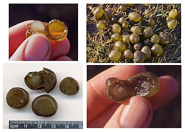
Fossils
Stromatolites are layered biochemical accretionary structures formed in shallow water by the trapping, binding, and cementation of sedimentary grains by biofilms (microbial mats) of microorganisms, especially cyanobacteria.[150]
During the Precambrian, stromatolite communities of microorganisms grew in most marine and non-marine environments in the photic zone. After the Cambrian explosion of marine animals, grazing on the stromatolite mats by herbivores greatly reduced the occurrence of the stromatolites in marine environments. Since then, they are found mostly in hypersaline conditions where grazing invertebrates cannot live (e.g. Shark Bay, Western Australia). Stromatolites provide ancient records of life on Earth by fossil remains which date from 3.5 Ga ago.[151] As of 2010[update] the oldest undisputed evidence of cyanobacteria is from 2.1 Ga ago, but there is some evidence for them as far back as 2.7 Ga ago. Oxygen concentrations in the atmosphere remained around or below 1% of today's level until 2.4 Ga ago (the Great Oxygenation Event). The rise in oxygen may have caused a fall in the concentration of atmospheric methane, and triggered the Huronian glaciation from around 2.4 to 2.1 Ga ago. In this way, cyanobacteria may have killed off much of the other bacteria of the time.[152]
Oncolites are sedimentary structures composed of oncoids, which are layered structures formed by cyanobacterial growth. Oncolites are similar to stromatolites, but instead of forming columns, they form approximately spherical structures that were not attached to the underlying substrate as they formed.[153] The oncoids often form around a central nucleus, such as a shell fragment,[154] and a calcium carbonate structure is deposited by encrusting microbes. Oncolites are indicators of warm waters in the photic zone, but are also known in contemporary freshwater environments.[155] These structures rarely exceed 10 cm in diameter.
One former classification scheme of cyanobacterial fossils divided them into the porostromata and the spongiostromata. These are now recognized as form taxa and considered taxonomically obsolete; however, some authors have advocated for the terms remaining informally to describe form and structure of bacterial fossils.[156]
-
 Stromatolites left behind by cyanobacteria are the oldest known fossils of life on Earth. This one-billion-year-old fossil is from Glacier National Park in Montana.
 -
 Oncolitic limestone formed from successive layers of calcium carbonate precipitated by cyanobacteria
 -
![Cyanobacterial remains of an annulated tubular microfossil Oscillatoriopsis longa [157] Scale bar: 100 μm](//upload.wikimedia.org/wikipedia/commons/thumb/3/38/Oscillatoriopsis_longa_fossil.jpg/270px-Oscillatoriopsis_longa_fossil.jpg) Cyanobacterial remains of an annulated tubular microfossil Oscillatoriopsis longa [157]
Cyanobacterial remains of an annulated tubular microfossil Oscillatoriopsis longa [157]
Scale bar: 100 μm

_3.jpg)
![Cyanobacterial remains of an annulated tubular microfossil Oscillatoriopsis longa [157] Scale bar: 100 μm](/wiki/File:Oscillatoriopsis_longa_fossil.jpg)
Classification

Historically, bacteria were first classified as plants constituting the class Schizomycetes, which along with the Schizophyceae (blue-green algae/Cyanobacteria) formed the phylum Schizophyta,[158] then in the phylum Monera in the kingdom Protista by Haeckel in 1866, comprising Protogens, Protamaeba, Vampyrella, Protomonae, and Vibrio, but not Nostoc and other cyanobacteria, which were classified with algae,[159] later reclassified as the Prokaryotes by Chatton.[160]
The cyanobacteria were traditionally classified by morphology into five sections, referred to by the numerals I–V. The first three – Chroococcales, Pleurocapsales, and Oscillatoriales – are not supported by phylogenetic studies. The latter two – Nostocales and Stigonematales – are monophyletic, and make up the heterocystous cyanobacteria.[161][162]
The members of Chroococales are unicellular and usually aggregate in colonies. The classic taxonomic criterion has been the cell morphology and the plane of cell division. In Pleurocapsales, the cells have the ability to form internal spores (baeocytes). The rest of the sections include filamentous species. In Oscillatoriales, the cells are uniseriately arranged and do not form specialized cells (akinetes and heterocysts).[163] In Nostocales and Stigonematales, the cells have the ability to develop heterocysts in certain conditions. Stigonematales, unlike Nostocales, include species with truly branched trichomes.[161]
Most taxa included in the phylum or division Cyanobacteria have not yet been validly published under The International Code of Nomenclature of Prokaryotes (ICNP) except:
- The classes Chroobacteria, Hormogoneae, and Gloeobacteria
- The orders Chroococcales, Gloeobacterales, Nostocales, Oscillatoriales, Pleurocapsales, and Stigonematales
- The families Prochloraceae and Prochlorotrichaceae
- The genera Halospirulina, Planktothricoides, Prochlorococcus, Prochloron, and Prochlorothrix
The remainder are validly published under the International Code of Nomenclature for algae, fungi, and plants.
Formerly, some bacteria, like Beggiatoa, were thought to be colorless Cyanobacteria.[164]
Relation to humans
Biotechnology and applications
The unicellular cyanobacterium Synechocystis sp. PCC6803 was the third prokaryote and first photosynthetic organism whose genome was completely sequenced.[165] It continues to be an important model organism.[166] Cyanothece ATCC 51142 is an important diazotrophic model organism. The smallest genomes have been found in Prochlorococcus spp. (1.7 Mb)[167][168] and the largest in Nostoc punctiforme (9 Mb).[169] Those of Calothrix spp. are estimated at 12–15 Mb,[170] as large as yeast.

Recent research has suggested the potential application of cyanobacteria to the generation of renewable energy by directly converting sunlight into electricity. Internal photosynthetic pathways can be coupled to chemical mediators that transfer electrons to external electrodes.[171] In the shorter term, efforts are underway to commercialize algae-based fuels such as diesel, gasoline, and jet fuel.[44][172][173]

Researchers from a company called Algenol have cultured genetically modified cyanobacteria in sea water inside a clear plastic enclosure so they first make sugar (pyruvate) from CO2 and the water via photosynthesis. Then, the bacteria secrete ethanol from the cell into the salt water. As the day progresses, and the solar radiation intensifies, ethanol concentrations build up and the ethanol itself evaporates onto the roof of the enclosure. As the sun recedes, evaporated ethanol and water condense into droplets, which run along the plastic walls and into ethanol collectors, from where it is extracted from the enclosure with the water and ethanol separated outside the enclosure. As of March 2013, Algenol was claiming to have tested its technology in Florida and to have achieved yields of 9,000 US gallons per acre per year.[174] This could potentially meet US demands for ethanol in gasoline in 2025, assuming a B30 blend, from an area of around half the size of California's San Bernardino County, requiring less than one-tenth of the area than ethanol from other biomass, such as corn, and only very limited amounts of fresh water.[175]
Cyanobacteria may possess the ability to produce substances that could one day serve as anti-inflammatory agents and combat bacterial infections in humans.[176] Cyanobacteria's photosynthetic output of sugar and oxygen has been demonstrated to have therapeutic value in rats with heart attacks.[177] While cyanobacteria can naturally produce various secondary metabolites, they can serve as advantageous hosts for plant-derived metabolites production owing to biotechnological advances in systems biology and synthetic biology.[178]
Spirulina's extracted blue color is used as a natural food coloring.[179]
Researchers from several space agencies argue that cyanobacteria could be used for producing goods for human consumption in future manned outposts on Mars, by transforming materials available on this planet.[180]
Rich source of human nutrition

Some cyanobacteria are sold as food, notably Arthrospira platensis (Spirulina) and others (Aphanizomenon flos-aquae) .[181]
Some microalgae contain substances of high biological value, such as polyunsaturated fatty acids, amino acids, proteins, pigments, antioxidants, vitamins, and minerals.[182] Edible blue-green algae reduce the production of pro-inflammatory cytokines by inhibiting NF-κB pathway in macrophages and splenocytes.[183] Sulfate polysaccharides exhibit immunomodulatory, antitumor, antithrombotic, anticoagulant, anti-mutagenic, anti-inflammatory, antimicrobial, and even antiviral activity against HIV, herpes, and hepatitis.[184]
Health risks
Some cyanobacteria can produce neurotoxins, cytotoxins, endotoxins, and hepatotoxins (e.g., the microcystin-producing bacteria genus microcystis), which are collectively known as cyanotoxins.
Specific toxins include anatoxin-a, guanitoxin, aplysiatoxin, cyanopeptolin, cylindrospermopsin, domoic acid, nodularin R (from Nodularia), neosaxitoxin, and saxitoxin. Cyanobacteria reproduce explosively under certain conditions. This results in algal blooms which can become harmful to other species and pose a danger to humans and animals if the cyanobacteria involved produce toxins. Several cases of human poisoning have been documented, but a lack of knowledge prevents an accurate assessment of the risks,[185][186][187][188] and research by Linda Lawton, FRSE at Robert Gordon University, Aberdeen and collaborators has 30 years of examining the phenomenon and methods of improving water safety.[189]
Recent studies suggest that significant exposure to high levels of cyanobacteria producing toxins such as BMAA can cause amyotrophic lateral sclerosis (ALS). People living within half a mile of cyanobacterially contaminated lakes have had a 2.3 times greater risk of developing ALS than the rest of the population; people around New Hampshire's Lake Mascoma had an up to 25 times greater risk of ALS than the expected incidence.[190] BMAA from desert crusts found throughout Qatar might have contributed to higher rates of ALS in Gulf War veterans.[186][191]
Chemical control
Several chemicals can eliminate cyanobacterial blooms from smaller water-based systems such as swimming pools. They include calcium hypochlorite, copper sulphate, cupricide, and simazine.[192] The calcium hypochlorite amount needed varies depending on the cyanobacteria bloom, and treatment is needed periodically. According to the Department of Agriculture Australia, a rate of 12 g of 70% material in 1000 l of water is often effective to treat a bloom.[192] Copper sulfate is also used commonly, but no longer recommended by the Australian Department of Agriculture, as it kills livestock, crustaceans, and fish.[192] Cupricide is a chelated copper product that eliminates blooms with lower toxicity risks than copper sulfate. Dosage recommendations vary from 190 ml to 4.8 l per 1000 m2.[192] Ferric alum treatments at the rate of 50 mg/l will reduce algae blooms.[192][193] Simazine, which is also a herbicide, will continue to kill blooms for several days after an application. Simazine is marketed at different strengths (25, 50, and 90%), the recommended amount needed for one cubic meter of water per product is 25% product 8 ml; 50% product 4 ml; or 90% product 2.2 ml.[192]
Climate change
Climate change is likely to increase the frequency, intensity and duration of cyanobacterial blooms in many eutrophic lakes, reservoirs and estuaries.[194][38] Bloom-forming cyanobacteria produce a variety of neurotoxins, hepatotoxins and dermatoxins, which can be fatal to birds and mammals (including waterfowl, cattle and dogs) and threaten the use of waters for recreation, drinking water production, agricultural irrigation and fisheries.[38] Toxic cyanobacteria have caused major water quality problems, for example in Lake Taihu (China), Lake Erie (USA), Lake Okeechobee (USA), Lake Victoria (Africa) and the Baltic Sea.[38][195][196][197]
Climate change favours cyanobacterial blooms both directly and indirectly.[38] Many bloom-forming cyanobacteria can grow at relatively high temperatures.[198] Increased thermal stratification of lakes and reservoirs enables buoyant cyanobacteria to float upwards and form dense surface blooms, which gives them better access to light and hence a selective advantage over nonbuoyant phytoplankton organisms.[199][200] Protracted droughts during summer increase water residence times in reservoirs, rivers and estuaries, and these stagnant warm waters can provide ideal conditions for cyanobacterial bloom development.[201][197]
The capacity of the harmful cyanobacterial genus Microcystis to adapt to elevated CO2 levels was demonstrated in both laboratory and field experiments.[202] Microcystis spp. take up CO2 and HCO3− and accumulate inorganic carbon in carboxysomes, and strain competitiveness was found to depend on the concentration of inorganic carbon. As a result, climate change and increased CO2 levels are expected to affect the strain composition of cyanobacterial blooms.[202][197]
Gallery
-
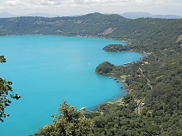 Cyanobacteria activity turns Coatepeque Caldera lake a turquoise color
 -
 Cyanobacterial bloom near Fiji
 -
 Cyanobacteria in Lake Köyliö.
 -
Video – Oscillatoria and Gleocapsa – with oscillatory movement as filaments of Oscillatoria orient towards light


See also
- Archean Eon
- Bacterial phyla, other major lineages of Bacteria
- Biodiesel
- Cyanobiont
- Endosymbiotic theory
- Geological history of oxygen
- Hypolith
References
- ^ "Cyanophyceae" (Document). Access Science. doi:10.1036/1097-8542.175300.
{{cite document}}: Unknown parameter|access-date=ignored (help); Unknown parameter|url=ignored (help) - ^ Oren A (September 2004). "A proposal for further integration of the cyanobacteria under the Bacteriological Code". International Journal of Systematic and Evolutionary Microbiology. 54 (Pt 5): 1895–902. doi:10.1099/ijs.0.03008-0. PMID 15388760.
- ^ Komárek J, Kaštovský J, Mareš J, Johansen JR (2014). "Taxonomic classification of cyanoprokaryotes (cyanobacterial genera) 2014, using a polyphasic approach" (PDF). Preslia. 86: 295–335.
- ^ Sinha, Rajeshwar P.; Häder, Donat-P. (2008). "UV-protectants in cyanobacteria" (PDF). Plant Science. 174 (3): 278–289. doi:10.1016/j.plantsci.2007.12.004. Archived from the original (PDF) on 15 April 2021.
- ^ "cyan | Origin and meaning of cyan by Online Etymology Dictionary". www.etymonline.com. Retrieved 21 January 2018.
- ^ "Henry George Liddell, Robert Scott, A Greek-English Lexicon, κύα^νος". www.perseus.tufts.edu. Retrieved 21 January 2018.
- ^ "Life History and Ecology of Cyanobacteria". University of California Museum of Paleontology. Archived from the original on 19 September 2012. Retrieved 17 July 2012.
- ^ "Taxonomy Browser – Cyanobacteria". National Center for Biotechnology Information. NCBI:txid1117. Retrieved 12 April 2018.
- ^ Allaby, M, ed. (1992). "Algae". The Concise Dictionary of Botany. Oxford: Oxford University Press.
- ^ Stal LJ, Cretoiu MS (2016). The Marine Microbiome: An Untapped Source of Biodiversity and Biotechnological Potential. Springer. ISBN 978-3319330006.
- ^ Monchamp, Marie-Eve; Spaak, Piet; Pomati, Francesco (27 July 2019). "Long Term Diversity and Distribution of Non-photosynthetic Cyanobacteria in Peri-Alpine Lakes". Frontiers in Microbiology. 0. doi:10.3389/fmicb.2018.03344 – via Frontiers.
{{cite journal}}: CS1 maint: unflagged free DOI (link) - ^ Liberton M, Pakrasi HB (2008). "Chapter 10. Membrane Systems in Cyanobacteria". In Herrero A, Flore E (eds.). The Cyanobacteria: Molecular Biology, Genomics, and Evolution. Norwich, United Kingdom: Horizon Scientific Press. pp. 217–87. ISBN 978-1-904455-15-8.
- ^ Liberton M, Page LE, O'Dell WB, O'Neill H, Mamontov E, Urban VS, Pakrasi HB (February 2013). "Organization and flexibility of cyanobacterial thylakoid membranes examined by neutron scattering". The Journal of Biological Chemistry. 288 (5): 3632–40. doi:10.1074/jbc.M112.416933. PMC 3561581. PMID 23255600.
{{cite journal}}: CS1 maint: unflagged free DOI (link) - ^ Whitton BA, ed. (2012). "The fossil record of cyanobacteria". Ecology of Cyanobacteria II: Their Diversity in Space and Time. Springer Science & Business Media. pp. 17–. ISBN 978-94-007-3855-3.
- ^ Basic Biology (18 March 2016). "Bacteria".
- ^ Dodds WK, Gudder DA, Mollenhauer D (1995). "The ecology of 'Nostoc'". Journal of Phycology. 31: 2–18. doi:10.1111/j.0022-3646.1995.00002.x. S2CID 85011483.
- ^ Meeks JC, Elhai J, Thiel T, Potts M, Larimer F, Lamerdin J, Predki P, Atlas R (2001). "An overview of the genome of Nostoc punctiforme, a multicellular, symbiotic cyanobacterium". Photosynthesis Research. 70 (1): 85–106. doi:10.1023/A:1013840025518. PMID 16228364. S2CID 8752382.
- ^ a b Golden JW, Yoon HS (December 1998). "Heterocyst formation in Anabaena". Current Opinion in Microbiology. 1 (6): 623–9. doi:10.1016/s1369-5274(98)80106-9. PMID 10066546.
- ^ a b c Fay P (June 1992). "Oxygen relations of nitrogen fixation in cyanobacteria". Microbiological Reviews. 56 (2): 340–73. doi:10.1128/MMBR.56.2.340-373.1992. PMC 372871. PMID 1620069.
- ^ a b Claessen, Dennis; Rozen, Daniel E.; Kuipers, Oscar P.; Søgaard-Andersen, Lotte; Van Wezel, Gilles P. (2014). "Bacterial solutions to multicellularity: A tale of biofilms, filaments and fruiting bodies". Nature Reviews Microbiology. 12 (2): 115–124. doi:10.1038/nrmicro3178. hdl:11370/0db66a9c-72ef-4e11-a75d-9d1e5827573d. PMID 24384602. S2CID 20154495.
- ^ Nürnberg, Dennis J.; Mariscal, Vicente; Parker, Jamie; Mastroianni, Giulia; Flores, Enrique; Mullineaux, Conrad W. (2014). "Branching and intercellular communication in the Section V cyanobacterium Mastigocladus laminosus, a complex multicellular prokaryote". Molecular Microbiology. 91 (5): 935–949. doi:10.1111/mmi.12506. PMID 24383541. S2CID 25479970.
- ^ Herrero, Antonia; Stavans, Joel; Flores, Enrique (2016). "The multicellular nature of filamentous heterocyst-forming cyanobacteria". FEMS Microbiology Reviews. 40 (6): 831–854. doi:10.1093/femsre/fuw029. PMID 28204529.
- ^ a b c d e f Aguilera, Anabella; Klemenčič, Marina; Sueldo, Daniela J.; Rzymski, Piotr; Giannuzzi, Leda; Martin, María Victoria (2021). "Cell Death in Cyanobacteria: Current Understanding and Recommendations for a Consensus on Its Nomenclature". Frontiers in Microbiology. 12: 631654. doi:10.3389/fmicb.2021.631654. PMC 7965980. PMID 33746925.
 Material was copied from this source, which is available under a Creative Commons Attribution 4.0 International License.
- ^ Risser DD, Chew WG, Meeks JC (April 2014). "Genetic characterization of the hmp locus, a chemotaxis-like gene cluster that regulates hormogonium development and motility in Nostoc punctiforme". Molecular Microbiology. 92 (2): 222–33. doi:10.1111/mmi.12552. PMID 24533832. S2CID 37479716.
- ^ Khayatan B, Bains DK, Cheng MH, Cho YW, Huynh J, Kim R, Omoruyi OH, Pantoja AP, Park JS, Peng JK, Splitt SD, Tian MY, Risser DD (May 2017). "A Putative O-Linked β-N-Acetylglucosamine Transferase Is Essential for Hormogonium Development and Motility in the Filamentous Cyanobacterium Nostoc punctiforme". Journal of Bacteriology. 199 (9): e00075–17. doi:10.1128/JB.00075-17. PMC 5388816. PMID 28242721.
- ^ Singh. Text Book of Botany Diversity of Microbes And Cryptogams. Rastogi Publications. ISBN 978-8171338894.
- ^ "Differences between Bacteria and Cyanobacteria". Microbiology Notes. 29 October 2015. Retrieved 21 January 2018.
- ^ Walsby AE (March 1994). "Gas vesicles". Microbiological Reviews. 58 (1): 94–144. doi:10.1128/MMBR.58.1.94-144.1994. PMC 372955. PMID 8177173.
- ^ Sims GK, Dunigan EP (1984). "Diurnal and seasonal variations in nitrogenase activity (C2H2 reduction) of rice roots". Soil Biology and Biochemistry. 16: 15–18. doi:10.1016/0038-0717(84)90118-4.
- ^ Bocchi S, Malgioglio A (2010). "Azolla-Anabaena as a Biofertilizer for Rice Paddy Fields in the Po Valley, a Temperate Rice Area in Northern Italy". International Journal of Agronomy. 2010: 1–5. doi:10.1155/2010/152158.
- ^ Esteves-Ferreira, Alberto A.; Cavalcanti, João Henrique Frota; Vaz, Marcelo Gomes Marçal Vieira; Alvarenga, Luna V.; Nunes-Nesi, Adriano; Araújo, Wagner L. (2017). "Cyanobacterial nitrogenases: Phylogenetic diversity, regulation and functional predictions". Genetics and Molecular Biology. 40 (1 suppl 1): 261–275. doi:10.1590/1678-4685-GMB-2016-0050. PMC 5452144. PMID 28323299.
- ^ Whitton, Brian A. (5 July 2012). Ecology of Cyanobacteria II: Their Diversity in Space and Time. ISBN 9789400738553.
- ^ Schopf, J.; Packer, B. (1987). "Early Archean (3.3-billion to 3.5-billion-year-old) microfossils from Warrawoona Group, Australia". Science. 237 (4810): 70–73. Bibcode:1987Sci...237...70S. doi:10.1126/science.11539686. PMID 11539686.
- ^ Bullerjahn, George S.; Post, Anton F. (2014). "Physiology and molecular biology of aquatic cyanobacteria". Frontiers in Microbiology. 5: 359. doi:10.3389/fmicb.2014.00359. PMC 4099938. PMID 25076944.
- ^ Tang, Weiyi; Wang, Seaver; Fonseca-Batista, Debany; Dehairs, Frank; Gifford, Scott; Gonzalez, Aridane G.; Gallinari, Morgane; Planquette, Hélène; Sarthou, Géraldine; Cassar, Nicolas (2019). "Revisiting the distribution of oceanic N2 fixation and estimating diazotrophic contribution to marine production". Nature Communications. 10 (1): 831. doi:10.1038/s41467-019-08640-0. PMC 6381160. PMID 30783106.
- ^ Bláha, Luděk; Babica, Pavel; Maršálek, Blahoslav (2009). "Toxins produced in cyanobacterial water blooms - toxicity and risks". Interdisciplinary Toxicology. 2 (2): 36–41. doi:10.2478/v10102-009-0006-2. PMC 2984099. PMID 21217843.
- ^ Paerl, Hans W.; Otten, Timothy G. (2013). "Harmful Cyanobacterial Blooms: Causes, Consequences, and Controls". Microbial Ecology. 65 (4): 995–1010. doi:10.1007/s00248-012-0159-y. PMID 23314096. S2CID 5718333.
- ^ a b c d e f Huisman, Jef; Codd, Geoffrey A.; Paerl, Hans W.; Ibelings, Bas W.; Verspagen, Jolanda M. H.; Visser, Petra M. (2018). "Cyanobacterial blooms". Nature Reviews Microbiology. 16 (8): 471–483. doi:10.1038/s41579-018-0040-1. PMID 29946124. S2CID 49427202.
- ^ Huokko, Tuomas; Ni, Tao; Dykes, Gregory F.; Simpson, Deborah M.; Brownridge, Philip; Conradi, Fabian D.; Beynon, Robert J.; Nixon, Peter J.; Mullineaux, Conrad W.; Zhang, Peijun; Liu, Lu-Ning (2021). "Probing the biogenesis pathway and dynamics of thylakoid membranes". Nature Communications. 12 (1): 3475. Bibcode:2021NatCo..12.3475H. doi:10.1038/s41467-021-23680-1. PMC 8190092. PMID 34108457.
- ^ Kerfeld CA, Heinhorst S, Cannon GC (2010). "Bacterial microcompartments". Annual Review of Microbiology. 64 (1): 391–408. doi:10.1146/annurev.micro.112408.134211. PMID 20825353.
- ^ Long BM, Badger MR, Whitney SM, Price GD (October 2007). "Analysis of carboxysomes from Synechococcus PCC7942 reveals multiple Rubisco complexes with carboxysomal proteins CcmM and CcaA". The Journal of Biological Chemistry. 282 (40): 29323–35. doi:10.1074/jbc.M703896200. PMID 17675289.
- ^ Vothknecht UC, Westhoff P (December 2001). "Biogenesis and origin of thylakoid membranes". Biochimica et Biophysica Acta (BBA) - Molecular Cell Research. 1541 (1–2): 91–101. doi:10.1016/S0167-4889(01)00153-7. PMID 11750665.
- ^ Sobiechowska-Sasim M, Stoń-Egiert J, Kosakowska A (February 2014). "Quantitative analysis of extracted phycobilin pigments in cyanobacteria – an assessment of spectrophotometric and spectrofluorometric methods". J Appl Phycol. 26 (5): 2065–74. doi:10.1007/s10811-014-0244-3. PMC 4200375. PMID 25346572.
- ^ a b Pisciotta JM, Zou Y, Baskakov IV (May 2010). Yang CH (ed.). "Light-dependent electrogenic activity of cyanobacteria". PLOS ONE. 5 (5): e10821. Bibcode:2010PLoSO...510821P. doi:10.1371/journal.pone.0010821. PMC 2876029. PMID 20520829.
{{cite journal}}: CS1 maint: unflagged free DOI (link) - ^ a b c Vermaas WF (2001). "Photosynthesis and Respiration in Cyanobacteria". Photosynthesis and Respiration in Cyanobacteria. eLS. John Wiley & Sons, Ltd. doi:10.1038/npg.els.0001670. ISBN 978-0-470-01590-2. S2CID 19016706.
- ^ Armstronf JE (2015). How the Earth Turned Green: A Brief 3.8-Billion-Year History of Plants. The University of Chicago Press. ISBN 978-0-226-06977-7.
- ^ Klatt JM, de Beer D, Häusler S, Polerecky L (2016). "Cyanobacteria in Sulfidic Spring Microbial Mats Can Perform Oxygenic and Anoxygenic Photosynthesis Simultaneously during an Entire Diurnal Period". Frontiers in Microbiology. 7: 1973. doi:10.3389/fmicb.2016.01973. PMC 5156726. PMID 28018309.
{{cite journal}}: CS1 maint: unflagged free DOI (link) - ^ Grossman AR, Schaefer MR, Chiang GG, Collier JL (September 1993). "The phycobilisome, a light-harvesting complex responsive to environmental conditions". Microbiological Reviews. 57 (3): 725–49. doi:10.1128/MMBR.57.3.725-749.1993. PMC 372933. PMID 8246846.
- ^ "Colors from bacteria | Causes of Color". www.webexhibits.org. Retrieved 22 January 2018.
- ^ Garcia-Pichel F (2009). "Cyanobacteria". In Schaechter M (ed.). Encyclopedia of Microbiology (third ed.). pp. 107–24. doi:10.1016/B978-012373944-5.00250-9. ISBN 978-0-12-373944-5.
- ^ Kehoe DM (May 2010). "Chromatic adaptation and the evolution of light color sensing in cyanobacteria". Proceedings of the National Academy of Sciences of the United States of America. 107 (20): 9029–30. Bibcode:2010PNAS..107.9029K. doi:10.1073/pnas.1004510107. PMC 2889117. PMID 20457899.
- ^ Kehoe DM, Gutu A (2006). "Responding to color: the regulation of complementary chromatic adaptation". Annual Review of Plant Biology. 57: 127–50. doi:10.1146/annurev.arplant.57.032905.105215. PMID 16669758.
- ^ Palenik B, Haselkorn R (January 1992). "Multiple evolutionary origins of prochlorophytes, the chlorophyll b-containing prokaryotes". Nature. 355 (6357): 265–67. Bibcode:1992Natur.355..265P. doi:10.1038/355265a0. PMID 1731224. S2CID 4244829.
- ^ Urbach E, Robertson DL, Chisholm SW (January 1992). "Multiple evolutionary origins of prochlorophytes within the cyanobacterial radiation". Nature. 355 (6357): 267–70. Bibcode:1992Natur.355..267U. doi:10.1038/355267a0. PMID 1731225. S2CID 2011379.
- ^ Cohen Y, Jørgensen BB, Revsbech NP, Poplawski R (February 1986). "Adaptation to Hydrogen Sulfide of Oxygenic and Anoxygenic Photosynthesis among Cyanobacteria". Applied and Environmental Microbiology. 51 (2): 398–407. doi:10.1128/AEM.51.2.398-407.1986. PMC 238881. PMID 16346996.
- ^ Blankenship RE (2014). Molecular Mechanisms of Photosynthesis. Wiley-Blackwell. pp. 147–73. ISBN 978-1-4051-8975-0.
- ^ Och LM, Shields-Zhou GA (January 2012). "The Neoproterozoic oxygenation event: Environmental perturbations and biogeochemical cycling". Earth-Science Reviews. 110 (1–4): 26–57. Bibcode:2012ESRv..110...26O. doi:10.1016/j.earscirev.2011.09.004.
- ^ Adams DG, Bergman B, Nierzwicki-Bauer SA, Duggan PS, Rai AN, Schüßler A (2013). The Prokaryotes. Springer, Berlin, Heidelberg. pp. 359–400. doi:10.1007/978-3-642-30194-0_17. ISBN 978-3-642-30193-3.
- ^ Smith A (1973). "Synthesis of metabolic intermediates". In Carr NG, Whitton BA (eds.). The Biology of Blue-green Algae. University of California Press. pp. 30–. ISBN 978-0-520-02344-4.
- ^ Jangoux M (1987). "Diseases of Echinodermata. I. Agents microorganisms and protistans". Dis. Aquat. Org. 2: 147–62. doi:10.3354/dao002147.
- ^ Kinne O, ed. (1980). Diseases of Marine Animals (PDF). Vol. 1. Chichester, UK: John Wiley & Sons. ISBN 978-0-471-99584-5.
- ^ Kristiansen A (1964). "Sarcinastrum urosporae, a Colourless Parasitic Blue-green Alga" (PDF). Phycologia. 4 (1): 19–22. doi:10.2216/i0031-8884-4-1-19.1. Archived from the original (PDF) on 6 January 2015.
- ^ a b Keeling PJ (2013). "The number, speed, and impact of plastid endosymbioses in eukaryotic evolution". Annual Review of Plant Biology. 64: 583–607. doi:10.1146/annurev-arplant-050312-120144. PMID 23451781. S2CID 207679266.
- ^ Moore KR, Magnabosco C, Momper L, Gold DA, Bosak T, Fournier GP (2019). "An Expanded Ribosomal Phylogeny of Cyanobacteria Supports a Deep Placement of Plastids". Frontiers in Microbiology. 10: 1612. doi:10.3389/fmicb.2019.01612. PMC 6640209. PMID 31354692.
{{cite journal}}: CS1 maint: unflagged free DOI (link) - ^ Howe CJ, Barbrook AC, Nisbet RE, Lockhart PJ, Larkum AW (August 2008). "The origin of plastids". Philosophical Transactions of the Royal Society of London. Series B, Biological Sciences. 363 (1504): 2675–85. doi:10.1098/rstb.2008.0050. PMC 2606771. PMID 18468982.
- ^ a b Rodríguez-Ezpeleta N, Brinkmann H, Burey SC, Roure B, Burger G, Löffelhardt W, Bohnert HJ, Philippe H, Lang BF (July 2005). "Monophyly of primary photosynthetic eukaryotes: green plants, red algae, and glaucophytes". Current Biology. 15 (14): 1325–30. doi:10.1016/j.cub.2005.06.040. PMID 16051178.
- ^ Adl SM, Simpson AG, Lane CE, Lukeš J, Bass D, Bowser SS, Brown MW, Burki F, Dunthorn M, Hampl V, Heiss A, Hoppenrath M, Lara E, Le Gall L, Lynn DH, McManus H, Mitchell EA, Mozley-Stanridge SE, Parfrey LW, Pawlowski J, Rueckert S, Shadwick L, Shadwick L, Schoch CL, Smirnov A, Spiegel FW (September 2012). "The revised classification of eukaryotes". The Journal of Eukaryotic Microbiology. 59 (5): 429–93. doi:10.1111/j.1550-7408.2012.00644.x. PMC 3483872. PMID 23020233.
- ^ Price DC, Chan CX, Yoon HS, Yang EC, Qiu H, Weber AP, Schwacke R, Gross J, Blouin NA, Lane C, Reyes-Prieto A, Durnford DG, Neilson JA, Lang BF, Burger G, Steiner JM, Löffelhardt W, Meuser JE, Posewitz MC, Ball S, Arias MC, Henrissat B, Coutinho PM, Rensing SA, Symeonidi A, Doddapaneni H, Green BR, Rajah VD, Boore J, Bhattacharya D (February 2012). "Cyanophora paradoxa genome elucidates origin of photosynthesis in algae and plants". Science. 335 (6070): 843–47. Bibcode:2012Sci...335..843P. doi:10.1126/science.1213561. PMID 22344442. S2CID 17190180.
- ^ a b Ponce-Toledo RI, Deschamps P, López-García P, Zivanovic Y, Benzerara K, Moreira D (February 2017). "An Early-Branching Freshwater Cyanobacterium at the Origin of Plastids". Current Biology. 27 (3): 386–91. doi:10.1016/j.cub.2016.11.056. PMC 5650054. PMID 28132810.
- ^ Schimper, AF (1883). "Über die Entwicklung der Chlorophyllkörner und Farbkörper" [About the development of the chlorophyll grains and stains]. Bot. Zeitung (in German). 41: 105–14, 121–31, 137–46, 153–62. Archived from the original on 19 October 2013.
- ^ Alberts B (2002). Molecular biology of the cell (4. ed.). New York [u.a.]: Garland. ISBN 978-0-8153-4072-0.
- ^ Mereschkowsky C (1905). "Über Natur und Ursprung der Chromatophoren im Pflanzenreiche" [About the nature and origin of chromatophores in the vegetable kingdom]. Biol Centralbl (in German). 25: 593–604.
- ^ Sagan L (March 1967). "On the origin of mitosing cells". Journal of Theoretical Biology. 14 (3): 255–74. doi:10.1016/0022-5193(67)90079-3. PMID 11541392.
- ^ Schwartz RM, Dayhoff MO (January 1978). "Origins of prokaryotes, eukaryotes, mitochondria, and chloroplasts". Science. 199 (4327): 395–403. Bibcode:1978Sci...199..395S. doi:10.1126/science.202030. PMID 202030.
- ^ Archibald JM (August 2015). "Genomic perspectives on the birth and spread of plastids". Proceedings of the National Academy of Sciences of the United States of America. 112 (33): 10147–53. Bibcode:2015PNAS..11210147A. doi:10.1073/pnas.1421374112. PMC 4547232. PMID 25902528.
- ^ Blankenship RE (October 2010). "Early evolution of photosynthesis". Plant Physiology. 154 (2): 434–38. doi:10.1104/pp.110.161687. PMC 2949000. PMID 20921158.
- ^ Rockwell NC, Lagarias JC, Bhattacharya D (2014). "Primary endosymbiosis and the evolution of light and oxygen sensing in photosynthetic eukaryotes". Frontiers in Ecology and Evolution. 2 (66). doi:10.3389/fevo.2014.00066. PMC 4343542. PMID 25729749.
{{cite journal}}: CS1 maint: unflagged free DOI (link) - ^ Summarised in Cavalier-Smith T (April 2000). "Membrane heredity and early chloroplast evolution". Trends in Plant Science. 5 (4): 174–82. doi:10.1016/S1360-1385(00)01598-3. PMID 10740299.
- ^ Nowack EC, Melkonian M, Glöckner G (March 2008). "Chromatophore genome sequence of Paulinella sheds light on acquisition of photosynthesis by eukaryotes". Current Biology. 18 (6): 410–18. doi:10.1016/j.cub.2008.02.051. PMID 18356055.
- ^ Archibald JM (January 2009). "The puzzle of plastid evolution". Current Biology. 19 (2): R81–88. doi:10.1016/j.cub.2008.11.067. PMID 19174147.
- ^ "How do plants make oxygen? Ask cyanobacteria". Phys.org. Science X. 30 March 2017. Retrieved 26 October 2017.
- ^ Orkwiszewski KG, Kaney AR (June 1974). "Genetic transformation of the blue-green bacterium, Anacystis nidulans". Arch Mikrobiol. 98 (1): 31–37. doi:10.1007/BF00425265. PMID 4209657. S2CID 5635245.
- ^ Stevens SE, Porter RD (October 1980). "Transformation in Agmenellum quadruplicatum". Proc. Natl. Acad. Sci. U.S.A. 77 (10): 6052–56. Bibcode:1980PNAS...77.6052S. doi:10.1073/pnas.77.10.6052. PMC 350211. PMID 16592896.
- ^ Grigorieva G, Shestakov S (1982). "Transformation in the cyanobacterium Synechocystis sp. 6803". FEMS Microbiology Letters. 13 (4): 367–70. doi:10.1111/j.1574-6968.1982.tb08289.x.
- ^ Bernstein H, Bernstein C, Michod RE (January 2018). "Sex in microbial pathogens". Infect. Genet. Evol. 57: 8–25. doi:10.1016/j.meegid.2017.10.024. PMID 29111273.
- ^ a b Cassier-Chauvat C, Veaudor T, Chauvat F (2016). "Comparative Genomics of DNA Recombination and Repair in Cyanobacteria: Biotechnological Implications". Front Microbiol. 7: 1809. doi:10.3389/fmicb.2016.01809. PMC 5101192. PMID 27881980.
{{cite journal}}: CS1 maint: unflagged free DOI (link) - ^ Mazard S, Penesyan A, Ostrowski M, Paulsen IT, Egan S (2016). "Tiny microbes with a big impact: the role of cyanobacteria and their metabolites in shaping our future". Marine Drugs. 14 (5): 97. doi:10.3390/md14050097. PMC 4882571. PMID 27196915.
{{cite journal}}: CS1 maint: unflagged free DOI (link) - ^ de los Ríos A, Grube M, Sancho LG, Ascaso C (February 2007). "Ultrastructural and genetic characteristics of endolithic cyanobacterial biofilms colonizing Antarctic granite rocks". FEMS Microbiology Ecology. 59 (2): 386–95. doi:10.1111/j.1574-6941.2006.00256.x. PMID 17328119.
- ^ Vaughan T (2011). Mammalogy. Jones and Barlett. p. 21. ISBN 978-0763762995.
- ^ Schultz, Nora (30 August 2009). "Photosynthetic viruses keep world's oxygen levels up". New Scientist.
- ^ a b Jöhnk KD, Huisman J, Sharples J, Sommeijer B, Visser PM, Stroom JM (1 March 2008). "Summer heatwaves promote blooms of harmful cyanobacteria". Global Change Biology. 14 (3): 495–512. Bibcode:2008GCBio..14..495J. doi:10.1111/j.1365-2486.2007.01510.x.
- ^ "Linda Lawton – 11th International Conference on Toxic Cyanobacteria". Retrieved 25 June 2021.
- ^ Paerl HW, Paul VJ (April 2012). "Climate change: links to global expansion of harmful cyanobacteria". Water Research. 46 (5): 1349–63. doi:10.1016/j.watres.2011.08.002. PMID 21893330.
- ^ Thomas AD, Dougill AJ (15 March 2007). "Spatial and temporal distribution of cyanobacterial soil crusts in the Kalahari: Implications for soil surface properties". Geomorphology. 85 (1): 17–29. Bibcode:2007Geomo..85...17T. doi:10.1016/j.geomorph.2006.03.029.
- ^ Belnap J, Gardner JS (1993). "Soil Microstructure in Soils of the Colorado Plateau: The Role of the Cyanobacterium Microcoleus Vaginatus". The Great Basin Naturalist. 53 (1): 40–47. JSTOR 41712756.
- ^ Nadis S (December 2003). "The cells that rule the seas" (PDF). Scientific American. 289 (6): 52–53. Bibcode:2003SciAm.289f..52N. doi:10.1038/scientificamerican1203-52. PMID 14631732. Archived from the original (PDF) on 19 April 2014. Retrieved 19 April 2014.
- ^ Walsh PJ, Smith S, Fleming L, Solo-Gabriele H, Gerwick WH, eds. (2 September 2011). "Cyanobacteria and cyanobacterial toxins". Oceans and Human Health: Risks and Remedies from the Seas. Academic Press. pp. 271–96. ISBN 978-0-08-087782-2.
- ^ a b c d e f g Mullineaux, Conrad W.; Wilde, Annegret (2021). "The social life of cyanobacteria". eLife. 10. doi:10.7554/eLife.70327. PMC 8208810. PMID 34132636.
{{cite journal}}: CS1 maint: unflagged free DOI (link) Material was copied from this source, which is available under a Creative Commons Attribution 4.0 International License.
- ^ Kamennaya, Nina A.; Zemla, Marcin; Mahoney, Laura; Chen, Liang; Holman, Elizabeth; Holman, Hoi-Ying; Auer, Manfred; Ajo-Franklin, Caroline M.; Jansson, Christer (2018). "High pCO2-induced exopolysaccharide-rich ballasted aggregates of planktonic cyanobacteria could explain Paleoproterozoic carbon burial". Nature Communications. 9 (1): 2116. Bibcode:2018NatCo...9.2116K. doi:10.1038/s41467-018-04588-9. PMC 5974010. PMID 29844378.
- ^ a b Maeda, Kaisei; Okuda, Yukiko; Enomoto, Gen; Watanabe, Satoru; Ikeuchi, Masahiko (2021). "Biosynthesis of a sulfated exopolysaccharide, synechan, and bloom formation in the model cyanobacterium Synechocystis sp. Strain PCC 6803". eLife. 10. doi:10.7554/eLife.66538. PMC 8205485. PMID 34127188.
{{cite journal}}: CS1 maint: unflagged free DOI (link) - ^ a b Sim, Min Sub; Liang, Biqing; Petroff, Alexander P.; Evans, Alexander; Klepac-Ceraj, Vanja; Flannery, David T.; Walter, Malcolm R.; Bosak, Tanja (2012). "Oxygen-Dependent Morphogenesis of Modern Clumped Photosynthetic Mats and Implications for the Archean Stromatolite Record". Geosciences. 2 (4): 235–259. Bibcode:2012Geosc...2..235S. doi:10.3390/geosciences2040235. Material was copied from this source, which is available under a Creative Commons Attribution 3.0 International License.
- ^ a b Conradi, Fabian D.; Zhou, Rui-Qian; Oeser, Sabrina; Schuergers, Nils; Wilde, Annegret; Mullineaux, Conrad W. (2019). "Factors Controlling Floc Formation and Structure in the Cyanobacterium Synechocystis sp. Strain PCC 6803". Journal of Bacteriology. 201 (19). doi:10.1128/JB.00344-19. PMC 6755745. PMID 31262837.
- ^ Enomoto, Gen; Ikeuchi, Masahiko (2020). "Blue-/Green-Light-Responsive Cyanobacteriochromes Are Cell Shade Sensors in Red-Light Replete Niches". iScience. 23 (3): 100936. Bibcode:2020iSci...23j0936E. doi:10.1016/j.isci.2020.100936. PMC 7063230. PMID 32146329.
- ^ Galluzzi, L.; et al. (2015). "Essential versus accessory aspects of cell death: Recommendations of the NCCD 2015". Cell Death & Differentiation. 22 (1): 58–73. doi:10.1038/cdd.2014.137. PMC 4262782. PMID 25236395.
- ^ Allen, Rey; Rittmann, Bruce E.; Curtiss, Roy (2019). "Axenic Biofilm Formation and Aggregation by Synechocystis sp. Strain PCC 6803 Are Induced by Changes in Nutrient Concentration and Require Cell Surface Structures". Applied and Environmental Microbiology. 85 (7). doi:10.1128/AEM.02192-18. PMC 6585507. PMID 30709828.
- ^ Schuergers, Nils; Wilde, Annegret (2015). "Appendages of the Cyanobacterial Cell". Life. 5 (1): 700–715. doi:10.3390/life5010700. PMC 4390875. PMID 25749611.
- ^ a b Khayatan, Behzad; Meeks, John C.; Risser, Douglas D. (2015). "Evidence that a modified type IV pilus-like system powers gliding motility and polysaccharide secretion in filamentous cyanobacteria". Molecular Microbiology. 98 (6): 1021–1036. doi:10.1111/mmi.13205. PMID 26331359. S2CID 8749419.
- ^ Adams, David. W.; Stutzmann, Sandrine; Stoudmann, Candice; Blokesch, Melanie (2019). "DNA-uptake pili of Vibrio cholerae are required for chitin colonization and capable of kin recognition via sequence-specific self-interaction". Nature Microbiology. 4 (9): 1545–1557. doi:10.1038/s41564-019-0479-5. PMC 6708440. PMID 31182799.
- ^ Kromkamp, Jacco; Walsby, Anthony E. (1990). "A computer model of buoyancy and vertical migration in cyanobacteria". Journal of Plankton Research. 12: 161–183. doi:10.1093/plankt/12.1.161.
- ^ Aguilo-Ferretjans, Maria del Mar; Bosch, Rafael; Puxty, Richard J.; Latva, Mira; Zadjelovic, Vinko; Chhun, Audam; Sousoni, Despoina; Polin, Marco; Scanlan, David J.; Christie-Oleza, Joseph A. (2021). "Pili allow dominant marine cyanobacteria to avoid sinking and evade predation". Nature Communications. 12 (1): 1857. Bibcode:2021NatCo..12.1857A. doi:10.1038/s41467-021-22152-w. PMC 7994388. PMID 33767153.
- ^ Agustí, S. (2004). "Viability and niche segregation of Prochlorococcus and Synechococcus cells across the Central Atlantic Ocean." Accessed: 30 July 2021).
- ^ Agusti, Susana; Alou, EVA; Hoyer, Mark V.; Frazer, Thomas K.; Canfield, Daniel E. (2006). "Cell death in lake phytoplankton communities". Freshwater Biology. 51 (8): 1496–1506. doi:10.1111/j.1365-2427.2006.01584.x.
- ^ Franklin, Daniel J.; Brussaard, Corina P.D.; Berges, John A. (2006). "What is the role and nature of programmed cell death in phytoplankton ecology?". European Journal of Phycology. 41: 1–14. doi:10.1080/09670260500505433. S2CID 53599616.
- ^ Sigee, D. C.; Selwyn, A.; Gallois, P.; Dean, A. P. (2007). "Patterns of cell death in freshwater colonial cyanobacteria during the late summer bloom". Phycologia. 46 (3): 284–292. doi:10.2216/06-69.1. S2CID 86268392.
- ^ Berman-Frank, Ilana; Bidle, Kay D.; Haramaty, Liti; Falkowski, Paul G. (2004). "The demise of the marine cyanobacterium, Trichodesmium SPP., via an autocatalyzed cell death pathway". Limnology and Oceanography. 49 (4): 997–1005. Bibcode:2004LimOc..49..997B. doi:10.4319/lo.2004.49.4.0997.
- ^ Hu, Chenlin; Rzymski, Piotr (2019). "Programmed Cell Death-Like and Accompanying Release of Microcystin in Freshwater Bloom-Forming Cyanobacterium Microcystis: From Identification to Ecological Relevance". Toxins. 11 (12): 706. doi:10.3390/toxins11120706. PMC 6950475. PMID 31817272.
- ^ Meeks, John C.; Elhai, Jeff; Thiel, Teresa; Potts, Malcolm; Larimer, Frank; Lamerdin, Jane; Predki, Paul; Atlas, Ronald (2001). "An overview of the genome of Nostoc punctiforme, a multicellular, symbiotic cyanobacterium". Photosynthesis Research. 70 (1): 85–106. doi:10.1023/A:1013840025518. PMID 16228364. S2CID 8752382.
- ^ Duggan, Paula S.; Gottardello, Priscila; Adams, David G. (2007). "Molecular Analysis of Genes in Nostoc punctiforme Involved in Pilus Biogenesis and Plant Infection". Journal of Bacteriology. 189 (12): 4547–4551. doi:10.1128/JB.01927-06. PMC 1913353. PMID 17416648.
- ^ Khayatan, Behzad; Meeks, John C.; Risser, Douglas D. (2015). "Evidence that a modified type IV pilus-like system powers gliding motility and polysaccharide secretion in filamentous cyanobacteria". Molecular Microbiology. 98 (6): 1021–1036. doi:10.1111/mmi.13205. PMID 26331359. S2CID 8749419.
- ^ Wilde, Annegret; Mullineaux, Conrad W. (2015). "Motility in cyanobacteria: Polysaccharide tracks and Type IV pilus motors". Molecular Microbiology. 98 (6): 998–1001. doi:10.1111/mmi.13242. PMID 26447922. S2CID 22585994.
- ^ Waterbury, J. B.; Willey, J. M.; Franks, D. G.; Valois, F. W.; Watson, S. W. (1985). "A Cyanobacterium Capable of Swimming Motility". Science. 230 (4721): 74–76. Bibcode:1985Sci...230...74W. doi:10.1126/science.230.4721.74. PMID 17817167. S2CID 29180516.
- ^ Ehlers, Kurt; Oster, George (2012). "On the Mysterious Propulsion of Synechococcus". PLOS ONE. 7 (5): e36081. Bibcode:2012PLoSO...736081E. doi:10.1371/journal.pone.0036081. PMC 3342319. PMID 22567124.
{{cite journal}}: CS1 maint: unflagged free DOI (link) - ^ Miyata, Makoto; Robinson, Robert C.; Uyeda, Taro Q. P.; Fukumori, Yoshihiro; Fukushima, Shun‐Ichi; Haruta, Shin; Homma, Michio; Inaba, Kazuo; Ito, Masahiro; Kaito, Chikara; Kato, Kentaro; Kenri, Tsuyoshi; Kinosita, Yoshiaki; Kojima, Seiji; Minamino, Tohru; Mori, Hiroyuki; Nakamura, Shuichi; Nakane, Daisuke; Nakayama, Koji; Nishiyama, Masayoshi; Shibata, Satoshi; Shimabukuro, Katsuya; Tamakoshi, Masatada; Taoka, Azuma; Tashiro, Yosuke; Tulum, Isil; Wada, Hirofumi; Wakabayashi, Ken‐Ichi (2020). "Tree of motility – A proposed history of motility systems in the tree of life". Genes to Cells. 25 (1): 6–21. doi:10.1111/gtc.12737. PMC 7004002. PMID 31957229. Material was copied from this source, which is available under a Creative Commons Attribution 4.0 International License.
- ^ R. W. Castenholz (1982). "Motility and taxes". In N. G. Carr; B. A. Whitton (eds.). The biology of cyanobacteria. University of California Press, Berkeley and Los Angeles. pp. 413–439. ISBN 978-0-520-04717-4.
- ^ J. B. Waterbury; J. M. Willey; D. G. Franks; F. W. Valois & S. W. Watson (1985). "A cyanobacterium capable of swimming motility". Science. 230 (4721): 74–76. Bibcode:1985Sci...230...74W. doi:10.1126/science.230.4721.74. PMID 17817167.
- ^ a b c d e Sánchez-Baracaldo, Patricia (2015). "Origin of marine planktonic cyanobacteria". Scientific Reports. 5: 17418. Bibcode:2015NatSR...517418S. doi:10.1038/srep17418. PMC 4665016. PMID 26621203. Material was copied from this source, which is available under a Creative Commons Attribution 4.0 International License.
- ^ a b c Bekker, A.; Holland, H. D.; Wang, P.-L.; Rumble, D.; Stein, H. J.; Hannah, J. L.; Coetzee, L. L.; Beukes, N. J. (2004). "Dating the rise of atmospheric oxygen". Nature. 427 (6970): 117–120. Bibcode:2004Natur.427..117B. doi:10.1038/nature02260. PMID 14712267. S2CID 4329872.
- ^ Kump, L. R.; Junium, C.; Arthur, M. A.; Brasier, A.; Fallick, A.; Melezhik, V.; Lepland, A.; Ccrne, A. E.; Luo, G. (2011). "Isotopic Evidence for Massive Oxidation of Organic Matter Following the Great Oxidation Event". Science. 334 (6063): 1694–1696. Bibcode:2011Sci...334.1694K. doi:10.1126/science.1213999. PMID 22144465. S2CID 27680273.
- ^ Fralick, Philip; Davis, Don W.; Kissin, Stephen A. (2002). "The age of the Gunflint Formation, Ontario, Canada: Single zircon UPb age determinations from reworked volcanic ash". Canadian Journal of Earth Sciences. 39 (7): 1085–1091. Bibcode:2002CaJES..39.1085F. doi:10.1139/e02-028.
{{cite journal}}: C1 control character in|title=at position 68 (help) - ^ a b Holland, Heinrich D. (2006). "The oxygenation of the atmosphere and oceans". Philosophical Transactions of the Royal Society B: Biological Sciences. 361 (1470): 903–915. doi:10.1098/rstb.2006.1838. PMC 1578726. PMID 16754606.
- ^ a b c d Lyons, Timothy W.; Reinhard, Christopher T.; Planavsky, Noah J. (2014). "The rise of oxygen in Earth's early ocean and atmosphere". Nature. 506 (7488): 307–315. Bibcode:2014Natur.506..307L. doi:10.1038/nature13068. PMID 24553238. S2CID 4443958.
- ^ a b Sahoo, Swapan K.; Planavsky, Noah J.; Kendall, Brian; Wang, Xinqiang; Shi, Xiaoying; Scott, Clint; Anbar, Ariel D.; Lyons, Timothy W.; Jiang, Ganqing (2012). "Ocean oxygenation in the wake of the Marinoan glaciation". Nature. 489 (7417): 546–549. Bibcode:2012Natur.489..546S. doi:10.1038/nature11445. PMID 23018964. S2CID 4391882.
- ^ Och, Lawrence M.; Shields-Zhou, Graham A. (2012). "The Neoproterozoic oxygenation event: Environmental perturbations and biogeochemical cycling". Earth-Science Reviews. 110 (1–4): 26–57. Bibcode:2012ESRv..110...26O. doi:10.1016/j.earscirev.2011.09.004.
- ^ a b Canfield, D. E.; Poulton, S. W.; Narbonne, G. M. (2007). "Late-Neoproterozoic Deep-Ocean Oxygenation and the Rise of Animal Life". Science. 315 (5808): 92–95. Bibcode:2007Sci...315...92C. doi:10.1126/science.1135013. PMID 17158290. S2CID 24761414.
- ^ a b c Fike, D. A.; Grotzinger, J. P.; Pratt, L. M.; Summons, R. E. (2006). "Oxidation of the Ediacaran Ocean". Nature. 444 (7120): 744–747. Bibcode:2006Natur.444..744F. doi:10.1038/nature05345. PMID 17151665. S2CID 4337003.
- ^ Sánchez-Baracaldo, Patricia; Ridgwell, Andy; Raven, John A. (2014). "A Neoproterozoic Transition in the Marine Nitrogen Cycle". Current Biology. 24 (6): 652–657. doi:10.1016/j.cub.2014.01.041. PMID 24583016. S2CID 16756351.
- ^ Erwin, D. H.; Laflamme, M.; Tweedt, S. M.; Sperling, E. A.; Pisani, D.; Peterson, K. J. (2011). "The Cambrian Conundrum: Early Divergence and Later Ecological Success in the Early History of Animals". Science. 334 (6059): 1091–1097. Bibcode:2011Sci...334.1091E. doi:10.1126/science.1206375. PMID 22116879. S2CID 7737847.
- ^ Poulton, S. W.; Canfield, D. E. (2011). "Ferruginous Conditions: A Dominant Feature of the Ocean through Earth's History". Elements. 7 (2): 107–112. doi:10.2113/gselements.7.2.107.
- ^ Anbar, A. D.; Knoll, A. H. (2002). "Proterozoic Ocean Chemistry and Evolution: A Bioinorganic Bridge?". Science. 297 (5584): 1137–1142. Bibcode:2002Sci...297.1137A. doi:10.1126/science.1069651. PMID 12183619. S2CID 5578019.
- ^ Field, C. B.; Behrenfeld, M. J.; Randerson, J. T.; Falkowski, P. (1998). "Primary Production of the Biosphere: Integrating Terrestrial and Oceanic Components". Science. 281 (5374): 237–240. Bibcode:1998Sci...281..237F. doi:10.1126/science.281.5374.237. PMID 9657713.
- ^ a b Zehr, Jonathan P. (2011). "Nitrogen fixation by marine cyanobacteria". Trends in Microbiology. 19 (4): 162–173. doi:10.1016/j.tim.2010.12.004. PMID 21227699.
- ^ a b Thompson, A. W.; Foster, R. A.; Krupke, A.; Carter, B. J.; Musat, N.; Vaulot, D.; Kuypers, M. M. M.; Zehr, J. P. (2012). "Unicellular Cyanobacterium Symbiotic with a Single-Celled Eukaryotic Alga". Science. 337 (6101): 1546–1550. Bibcode:2012Sci...337.1546T. doi:10.1126/science.1222700. PMID 22997339. S2CID 7071725.
- ^ a b Johnson, Z. I.; Zinser, E. R.; Coe, A.; McNulty, N. P.; Woodward, E. M.; Chisholm, S. W. (2006). "Niche Partitioning Among Prochlorococcus Ecotypes Along Ocean-Scale Environmental Gradients". Science. 311 (5768): 1737–1740. Bibcode:2006Sci...311.1737J. doi:10.1126/science.1118052. PMID 16556835. S2CID 3549275.
- ^ a b Scanlan, D. J.; Ostrowski, M.; Mazard, S.; Dufresne, A.; Garczarek, L.; Hess, W. R.; Post, A. F.; Hagemann, M.; Paulsen, I.; Partensky, F. (2009). "Ecological Genomics of Marine Picocyanobacteria". Microbiology and Molecular Biology Reviews. 73 (2): 249–299. doi:10.1128/MMBR.00035-08. PMC 2698417. PMID 19487728.
- ^ Flombaum, P.; Gallegos, J. L.; Gordillo, R. A.; Rincon, J.; Zabala, L. L.; Jiao, N.; Karl, D. M.; Li, W. K. W.; Lomas, M. W.; Veneziano, D.; Vera, C. S.; Vrugt, J. A.; Martiny, A. C. (2013). "Present and future global distributions of the marine Cyanobacteria Prochlorococcus and Synechococcus". Proceedings of the National Academy of Sciences. 110 (24): 9824–9829. Bibcode:2013PNAS..110.9824F. doi:10.1073/pnas.1307701110. PMC 3683724. PMID 23703908.
- ^ Foster, Rachel A.; Kuypers, Marcel M M.; Vagner, Tomas; Paerl, Ryan W.; Musat, Niculina; Zehr, Jonathan P. (2011). "Nitrogen fixation and transfer in open ocean diatom–cyanobacterial symbioses". The ISME Journal. 5 (9): 1484–1493. doi:10.1038/ismej.2011.26. PMC 3160684. PMID 21451586.
- ^ Villareal, Tracy A. (1990). "Laboratory Culture and Preliminary Characterization of the Nitrogen-Fixing Rhizosolenia-Richelia Symbiosis". Marine Ecology. 11 (2): 117–132. Bibcode:1990MarEc..11..117V. doi:10.1111/j.1439-0485.1990.tb00233.x.
- ^ Janson; Wouters; Bergman; Carpenter (1999). "Host specificity in the Richelia-diatom symbiosis revealed by hetR gene sequence analysis". Environmental Microbiology. 1 (5): 431–438. doi:10.1046/j.1462-2920.1999.00053.x. PMID 11207763.
- ^ Riding, R. (2007). "The term stromatolite: towards an essential definition". Lethaia. 32 (4): 321–30. doi:10.1111/j.1502-3931.1999.tb00550.x.
- ^ Baumgartner RJ, et al. (2019). "Nano-porous pyrite and organic matter in 3.5-billion-year-old stromatolites record primordial life" (PDF). Geology. 47 (11): 1039–43. Bibcode:2019Geo....47.1039B. doi:10.1130/G46365.1.
- ^ Lane, Nick (6 February 2010) "First breath: Earth's billion-year struggle for oxygen". New Scientist, pp. 36–39. See accompanying graph as well.
- ^ Corsetti FA, Awramik SM, Pierce D (April 2003). "A complex microbiota from snowball Earth times: microfossils from the Neoproterozoic Kingston Peak Formation, Death Valley, USA". Proceedings of the National Academy of Sciences of the United States of America. 100 (8): 4399–404. Bibcode:2003PNAS..100.4399C. doi:10.1073/pnas.0730560100. PMC 153566. PMID 12682298.
- ^ Gutschick RC, Perry TG (1 November 1959). "Sappington (Kinderhookian) sponges and their environment [Montana]". Journal of Paleontology. 33 (6): 977–85. Retrieved 28 June 2007.
- ^ Riding, Robert. (1991). Calcareous Algae and Stromatolites, p. 32. Springer-Verlag Press.
- ^ Monty, C. L. (1981). Monty, Claude (ed.). "Spongiostromate vs. Porostromate Stromatolites and Oncolites". Phanerozoic Stromatolites. Berlin, Heidelberg: Springer: 1–4. doi:10.1007/978-3-642-67913-1_1. ISBN 978-3-642-67913-1.
- ^ Castellani, Christopher; Maas, Andreas; Eriksson, Mats E.; Haug, Joachim T.; Haug, Carolin; Waloszek, Dieter (2018). "First record of Cyanobacteria in Cambrian Orsten deposits of Sweden". Palaeontology. 61 (6): 855–880. doi:10.1111/pala.12374.
- ^ Von Nägeli C (1857). Caspary R (ed.). "Bericht über die Verhandlungen der 33. Versammlung deutscher Naturforscher und Ärzte, gehalten in Bonn von 18 bis 24 September 1857" [Report on the Proceedings of the 33rd Meeting of German Natural Scientists and Physicians, held in Bonn, 18 to 24 September 1857]. Botanische Zeitung. 15: 749–76.
- ^ Haeckel E (1867). Generelle Morphologie der Organismen. Reimer, Berlin.
- ^ Chatton É (1925). "Pansporella perplexa. Réflexions sur la biologie et la phylogénie des protozoaires". Ann. Sci. Nat. Zool. 10-VII: 1–84.
- ^ a b Gugger MF, Hoffmann L (March 2004). "Polyphyly of true branching cyanobacteria (Stigonematales)". International Journal of Systematic and Evolutionary Microbiology. 54 (Pt 2): 349–57. doi:10.1099/ijs.0.02744-0. PMID 15023942.
- ^ Howard-Azzeh M, Shamseer L, Schellhorn HE, Gupta RS (November 2014). "Phylogenetic analysis and molecular signatures defining a monophyletic clade of heterocystous cyanobacteria and identifying its closest relatives". Photosynthesis Research. 122 (2): 171–85. doi:10.1007/s11120-014-0020-x. PMID 24917519. S2CID 17745718.
- ^ Komárek J, Kaštovský J, Mareš J, Johansen JR (2014). "Taxonomic classification of cyanoprokaryotes (cyanobacterial genera) 2014, using a polyphasic approach" (PDF). Preslia. 86: 295–335.
- ^ Pringsheim EG (1963). Farblose Algen: Ein Beitrag zur Evolutionsforschung. Gustav Fischer Verlag.
- ^ Kaneko T, Sato S, Kotani H, Tanaka A, Asamizu E, Nakamura Y, Miyajima N, Hirosawa M, Sugiura M, Sasamoto S, Kimura T, Hosouchi T, Matsuno A, Muraki A, Nakazaki N, Naruo K, Okumura S, Shimpo S, Takeuchi C, Wada T, Watanabe A, Yamada M, Yasuda M, Tabata S (June 1996). "Sequence analysis of the genome of the unicellular cyanobacterium Synechocystis sp. strain PCC6803. II. Sequence determination of the entire genome and assignment of potential protein-coding regions". DNA Research. 3 (3): 109–36. doi:10.1093/dnares/3.3.109. PMID 8905231.
- ^ Tabei Y, Okada K, Tsuzuki M (April 2007). "Sll1330 controls the expression of glycolytic genes in Synechocystis sp. PCC 6803". Biochemical and Biophysical Research Communications. 355 (4): 1045–50. doi:10.1016/j.bbrc.2007.02.065. PMID 17331473.
- ^ Rocap G, Larimer FW, Lamerdin J, Malfatti S, Chain P, Ahlgren NA, Arellano A, Coleman M, Hauser L, Hess WR, Johnson ZI, Land M, Lindell D, Post AF, Regala W, Shah M, Shaw SL, Steglich C, Sullivan MB, Ting CS, Tolonen A, Webb EA, Zinser ER, Chisholm SW (August 2003). "Genome divergence in two Prochlorococcus ecotypes reflects oceanic niche differentiation". Nature. 424 (6952): 1042–47. Bibcode:2003Natur.424.1042R. doi:10.1038/nature01947. PMID 12917642. S2CID 4344597.
- ^ Dufresne A, Salanoubat M, Partensky F, Artiguenave F, Axmann IM, Barbe V, Duprat S, Galperin MY, Koonin EV, Le Gall F, Makarova KS, Ostrowski M, Oztas S, Robert C, Rogozin IB, Scanlan DJ, Tandeau de Marsac N, Weissenbach J, Wincker P, Wolf YI, Hess WR (August 2003). "Genome sequence of the cyanobacterium Prochlorococcus marinus SS120, a nearly minimal oxyphototrophic genome". Proceedings of the National Academy of Sciences of the United States of America. 100 (17): 10020–25. Bibcode:2003PNAS..10010020D. doi:10.1073/pnas.1733211100. PMC 187748. PMID 12917486.
- ^ Meeks JC, Elhai J, Thiel T, Potts M, Larimer F, Lamerdin J, Predki P, Atlas R (2001). "An overview of the genome of Nostoc punctiforme, a multicellular, symbiotic cyanobacterium". Photosynthesis Research. 70 (1): 85–106. doi:10.1023/A:1013840025518. PMID 16228364. S2CID 8752382.
- ^ Herdman M, Janvier M, Rippka R, Stanier RY (1979). "Genome Size of Cyanobacteria". Journal of General Microbiology. 111: 73–85. doi:10.1099/00221287-111-1-73.
- ^ Quintana N, Van der Kooy F, Van de Rhee MD, Voshol GP, Verpoorte R (August 2011). "Renewable energy from Cyanobacteria: energy production optimization by metabolic pathway engineering". Applied Microbiology and Biotechnology. 91 (3): 471–90. doi:10.1007/s00253-011-3394-0. PMC 3136707. PMID 21691792.
- ^ "Blue green bacteria may help generate 'green' electricity", The Hindu, 21 June 2010
- ^ "Joule wins key patent for GMO cyanobacteria that create fuels from sunlight, CO2, and water. Biofuels Digest (2010-09-14). Retrieved on 2011-04-06.
- ^ "Algenol Biofuels exceeds 9,000 gallons of ethanol per year per" (PDF). Algenol Biofuels. 6 March 2013. Archived from the original (PDF) on 20 April 2014.
- ^ Lane J (25 September 2012). "Take it to the Limit: Algenol and rising yields in advanced biofuels". Biofuels Digest.
- ^ Choi H, Mascuch SJ, Villa FA, Byrum T, Teasdale ME, Smith JE, et al. (May 2012). "Honaucins A-C, potent inhibitors of inflammation and bacterial quorum sensing: synthetic derivatives and structure-activity relationships". Chemistry & Biology. 19 (5): 589–98. doi:10.1016/j.chembiol.2012.03.014. PMC 3361693. PMID 22633410.
- ^ Frischkorn, Kyle (19 June 2017). "Need to Fix a Heart Attack? Try Photosynthesis". Smithsonian. Retrieved 20 May 2021.
- ^ Jeong, Yujin; Cho, Sang-Hyeok; Lee, Hookeun; Choi, Hyung-Kyoon; Kim, Dong-Myung; Lee, Choul-Gyun; Cho, Suhyung; Cho, Byung-Kwan (December 2020). "Current Status and Future Strategies to Increase Secondary Metabolite Production from Cyanobacteria". Microorganisms. 8 (12): 1849. doi:10.3390/microorganisms8121849. PMC 7761380. PMID 33255283.
{{cite journal}}: CS1 maint: unflagged free DOI (link) - ^ Newsome, Andrew G.; Culver, Catherine A.; van Breemen, Richard B. (16 July 2014). "Nature's Palette: The Search for Natural Blue Colorants". Journal of Agricultural and Food Chemistry. 62 (28): 6498–6511. doi:10.1021/jf501419q. ISSN 0021-8561.
- ^ Verseux C, Baqué M, Lehto K, de Vera JP, Rothschild LJ, Billi D (2016). "Sustainable life support on Mars – the potential roles of cyanobacteria". International Journal of Astrobiology. 15 (1): 65–92. Bibcode:2016IJAsB..15...65V. doi:10.1017/S147355041500021X.
- ^ Spolaore P, Joannis-Cassan C, Duran E, Isambert A (February 2006). "Commercial applications of microalgae". Journal of Bioscience and Bioengineering. 101 (2): 87–96. doi:10.1263/jbb.101.87. PMID 16569602. S2CID 16896655.
- ^ Christaki E, Florou-Paneri P, Bonos E (December 2011). "Microalgae: a novel ingredient in nutrition". International Journal of Food Sciences and Nutrition. 62 (8): 794–99. doi:10.3109/09637486.2011.582460. PMID 21574818. S2CID 45956239.
- ^ Ku CS, Pham TX, Park Y, Kim B, Shin MS, Kang I, Lee J (April 2013). "Edible blue-green algae reduce the production of pro-inflammatory cytokines by inhibiting NF-κB pathway in macrophages and splenocytes". Biochimica et Biophysica Acta (BBA) - General Subjects. 1830 (4): 2981–88. doi:10.1016/j.bbagen.2013.01.018. PMC 3594481. PMID 23357040.
- ^ Mišurcová L, Škrovánková S, Samek D, Ambrožová J, Machů L (2012). "Health benefits of algal polysaccharides in human nutrition". Advances in Food and Nutrition Research Volume 66. Vol. 66. pp. 75–145. doi:10.1016/B978-0-12-394597-6.00003-3. ISBN 978-0-12-394597-6. PMID 22909979.
{{cite book}}:|journal=ignored (help) - ^ Thébault L, Lesne J, Boutin JP (1995). "[Cyanobacteria, their toxins and health risks]". Médecine Tropicale. 55 (4): 375–80. PMID 8830224.
- ^ a b Blue-Green Algae (Cyanobacteria) and their Toxins. Hc-sc.gc.ca (2013-01-30). Retrieved on 2014-04-19.
- ^ Harmful Bloom in Lake Atitlán, Guatemala from NASA Earth Observatory, retrieved on 9 January 2010.
- ^ Weinrich C, Miller TR (2014). "Freshwater harmful algal blooms: toxins and children's health". Current Problems in Pediatrics and Adolescent Health Care. 44: 2–24.
- ^ "Professor Linda Lawton". rgu-repository.worktribe.com. Retrieved 25 June 2021.
- ^ Caller TA, Doolin JW, Haney JF, Murby AJ, West KG, Farrar HE, Ball A, Harris BT, Stommel EW (2009). "A cluster of amyotrophic lateral sclerosis in New Hampshire: a possible role for toxic cyanobacteria blooms". Amyotrophic Lateral Sclerosis. 10 Suppl 2: 101–08. doi:10.3109/17482960903278485. PMID 19929741. S2CID 35250897.
- ^ Cox PA, Richer R, Metcalf JS, Banack SA, Codd GA, Bradley WG (2009). "Cyanobacteria and BMAA exposure from desert dust: a possible link to sporadic ALS among Gulf War veterans". Amyotrophic Lateral Sclerosis. 10 Suppl 2: 109–17. doi:10.3109/17482960903286066. PMID 19929742. S2CID 1748627.
- ^ a b c d e f Main D (2006). "Toxic Algae Blooms" (PDF). Veterinary Pathologist, South Perth. agric.wa.gov.au. Retrieved 18 November 2014.
- ^ May V, Baker H (1978). "Reduction of toxic algae in farm dams by ferric alum". Techn. Bull. 19: 1–16.
- ^ Paerl, H. W.; Huisman, J. (2008). "CLIMATE: Blooms Like It Hot". Science. 320 (5872): 57–58. doi:10.1126/science.1155398. PMID 18388279. S2CID 142881074.
- ^ Sitoki, L.; Kurmayer, R.; Rott, E. (2012). "Spatial variation of phytoplankton composition, biovolume, and resulting microcystin concentrations in the Nyanza Gulf (Lake Victoria, Kenya)". Hydrobiologia. 691 (1): 109–122. doi:10.1007/s10750-012-1062-8. PMC 3968937. PMID 24683268.
- ^ Cox, Paul Alan; Brand, Larry E.; Murch, Susan J.; Tymm, Fiona J. M.; Powell, James T.; Banack, Sandra Anne; Metcalf, James S. (2018). "Public health responses to toxic cyanobacterial blooms: Perspectives from the 2016 Florida event". Water Policy. 20 (5): 919–932. doi:10.2166/wp.2018.012.
- ^ a b c Cavicchioli, Ricardo; Ripple, William J.; Timmis, Kenneth N.; Azam, Farooq; Bakken, Lars R.; Baylis, Matthew; Behrenfeld, Michael J.; Boetius, Antje; Boyd, Philip W.; Classen, Aimée T.; Crowther, Thomas W.; Danovaro, Roberto; Foreman, Christine M.; Huisman, Jef; Hutchins, David A.; Jansson, Janet K.; Karl, David M.; Koskella, Britt; Mark Welch, David B.; Martiny, Jennifer B. H.; Moran, Mary Ann; Orphan, Victoria J.; Reay, David S.; Remais, Justin V.; Rich, Virginia I.; Singh, Brajesh K.; Stein, Lisa Y.; Stewart, Frank J.; Sullivan, Matthew B.; et al. (2019). "Scientists' warning to humanity: Microorganisms and climate change". Nature Reviews Microbiology. 17 (9): 569–586. doi:10.1038/s41579-019-0222-5. PMC 7136171. PMID 31213707. Material was copied from this source, which is available under a Creative Commons Attribution 4.0 International License.
- ^ Visser, Petra M.; Verspagen, Jolanda M.H.; Sandrini, Giovanni; Stal, Lucas J.; Matthijs, Hans C.P.; Davis, Timothy W.; Paerl, Hans W.; Huisman, Jef (2016). "How rising CO2 and global warming may stimulate harmful cyanobacterial blooms". Harmful Algae. 54: 145–159. doi:10.1016/j.hal.2015.12.006. PMID 28073473.
- ^ Walsby, Anthony E.; Hayes, Paul K.; Boje, Rolf; Stal, Lucas J. (1997). "The selective advantage of buoyancy provided by gas vesicles for planktonic cyanobacteria in the Baltic Sea". New Phytologist. 136 (3): 407–417. doi:10.1046/j.1469-8137.1997.00754.x. PMID 33863010.
- ^ Jöhnk, Klaus D.; Huisman, JEF; Sharples, Jonathan; Sommeijer, BEN; Visser, Petra M.; Stroom, Jasper M. (2008). "Summer heatwaves promote blooms of harmful cyanobacteria". Global Change Biology. 14 (3): 495–512. Bibcode:2008GCBio..14..495J. doi:10.1111/j.1365-2486.2007.01510.x.
- ^ Lehman, P.W.; Kurobe, T.; Lesmeister, S.; Baxa, D.; Tung, A.; Teh, S.J. (2017). "Impacts of the 2014 severe drought on the Microcystis bloom in San Francisco Estuary". Harmful Algae. 63: 94–108. doi:10.1016/j.hal.2017.01.011. PMID 28366405.
- ^ a b Sandrini, Giovanni; Ji, Xing; Verspagen, Jolanda M. H.; Tann, Robert P.; Slot, Pieter C.; Luimstra, Veerle M.; Schuurmans, J. Merijn; Matthijs, Hans C. P.; Huisman, Jef (2016). "Rapid adaptation of harmful cyanobacteria to rising CO2". Proceedings of the National Academy of Sciences. 113 (33): 9315–9320. doi:10.1073/pnas.1602435113. PMC 4995953. PMID 27482094.
- Attribution
![]() This article incorporates text available under the CC BY 2.5 license.
This article incorporates text available under the CC BY 2.5 license.
Further reading
- Cribbs G (1997). Nature's Superfood: the Blue-Green Algae Revolution (first ed.). Newleaf. ISBN 978-0-7522-0569-4.
- Savage M (1994). The Millennial Project: Colonizing the Galaxy in Eight Easy Steps. Little Brown & Co. ISBN 978-0-316-77163-4.
- Fogg GE, Stewart WD, Fay P, Walsby AE (1973). The Blue-green Algae. London and New York: Academic Press. ISBN 978-0-12-261650-1.
- "Architects of the earth's atmosphere", Introduction to the Cyanobacteria, University of California, Berkeley, 3 February 2006.
- Whitton BA (2002). "Phylum Cyanophyta (Cyanobacteria)". The Freshwater Algal Flora of the British Isles. Cambridge: Cambridge University Press. ISBN 978-0-521-77051-4.
- Pentecost A, Franke U (2010). "Photosynthesis and calcification of the stromatolitic freshwater cyanobacterium Rivularia". Eur. J. Phycol. 45 (4): 345–53. doi:10.1080/09670262.2010.492914.
- Whitton BA, Potts M, eds. (2000). The Ecology of Cyanobacteria: their Diversity in Time and Space. Springer. ISBN 978-0-7923-4735-4.
- "From Micro-Algae to Blue Oil". ParisTech Review. December 2011. Archived from the original on 17 April 2016. Retrieved 2 March 2012.
External links
- What are Cyanobacteria and What are its Types?
- Webserver for Cyanobacteria Research
- Diving an Antarctic Time Capsule Filled With Primordial Life

| Groups |  | |
|---|---|---|
| Microbiology | ||
| Motion | ||
| Ecology | ||
| Plants | ||
| Marine | ||
| Human related | ||
| Techniques | ||
| Other | ||