
Epithelium: Difference between revisions
ClueBot NG (talk | contribs) m Reverting possible vandalism by 198.235.255.4 to version by Omnipaedista. False positive? Report it. Thanks, ClueBot NG. (1765141) (Bot) |
|||
| Line 9: | Line 9: | ||
===Basement membrane=== |
===Basement membrane=== |
||
All epithelial cells rest on a [[basement membrane]], which acts as a scaffolding on which epithelium can grow and regenerate after injuries.<ref>{{Cite book|author=McConnell, Thomas H.|title=The nature of disease: pathology for the health professions|publisher=Lippincott Williams & Wilkins|year=2006|isbn=978-0-7817-5317-3|page=55|url=http://books.google.com/books?id=chs_lilPFLwC&pg=PA55}}</ref> Epithelial tissue is [[innervated]], but [[avascular]]. This epithelial tissue must be nourished by substances diffusing from the blood vessels in the underlying tissue, but they don't have their own blood supply. The basement membrane acts as a selectively permeable membrane that determines which substances will be able to enter the epithelium.<ref name="Eurell-2006-p18"/><ref name="p. 3"/> |
All epithelial hey hay cells rest on a [[basement membrane]], which acts as a scaffolding on which epithelium can grow and regenerate after injuries.<ref>{{Cite book|author=McConnell, Thomas H.|title=The nature of disease: pathology for the health professions|publisher=Lippincott Williams & Wilkins|year=2006|isbn=978-0-7817-5317-3|page=55|url=http://books.google.com/books?id=chs_lilPFLwC&pg=PA55}}</ref> Epithelial tissue is [[innervated]], but [[avascular]]. This epithelial tissue must be nourished by substances diffusing from the blood vessels in the underlying tissue, but they don't have their own blood supply. The basement membrane acts as a selectively permeable membrane that determines which substances will be able to enter the epithelium.<ref name="Eurell-2006-p18"/><ref name="p. 3"/> |
||
===Cell junctions=== |
===Cell junctions=== |
||
Revision as of 17:33, 26 March 2014
| This article is part of a series on |
| Epithelia |
|---|
| Squamous epithelial cell |
| Columnar epithelial cell |
| Cuboidal epithelial cell |
| Specialised epithelia |
|
| Other |
Epithelium is one of the four basic types of animal tissue, along with connective tissue, muscle tissue and nervous tissue. Epithelial tissues line the cavities and surfaces of structures throughout the body, and also form many glands. Functions of epithelial cells include secretion, selective absorption, protection, transcellular transport and detection of sensation. In Greek ἐπί (epi) means "on" or "upon", and θηλή (thēlē) means "nipple".[1]
Epithelial layers are avascular, so they must receive nourishment via diffusion of substances from the underlying connective tissue, through the basement membrane.[2][3] Epithelia can also be organised into clusters of cells that function as exocrine and endocrine glands.
Structure
Cells in epithelium are very densely packed together like bricks in a wall, leaving very little intercellular space. The cells can form continuous sheets attached to each other at many locations by adherens junctions, tight junctions, and desmosomes.[4]
Basement membrane
All epithelial hey hay cells rest on a basement membrane, which acts as a scaffolding on which epithelium can grow and regenerate after injuries.[5] Epithelial tissue is innervated, but avascular. This epithelial tissue must be nourished by substances diffusing from the blood vessels in the underlying tissue, but they don't have their own blood supply. The basement membrane acts as a selectively permeable membrane that determines which substances will be able to enter the epithelium.[2][3]
Cell junctions
Cell junctions are especially abundant in epithelial tissues. They consist of protein complexes and provide contact between neighbouring cells, between a cell and the extracellular matrix, or they build up the paracellular barrier of epithelia and control the paracellular transport.[citation needed]
Cell junctions are the contact points between plasma membrane and tissue cells. There are mainly 5 different types of cell junctions. They are tight junctions, adherens junctions, desmosomes, hemidesmosomes, and gap junctions. Tight junctions are a pair of trans-membrane protein fused on outer plasma membrane. Adherens junctions are a plaque (protein layer on the inside plasma membrane) which attaches both cells' microfilaments. Desmosomes attach to the microfilaments of cytoskeleton made up of keratin protein. Hemidesmosomes resemble desmosomes on a section. They are made up of the integrin (a transmembraner protein) instead of cadherin. They attach the epithelial cell to the basement membrane. Gap junctions connect the cytoplasm of two cells and are made up of proteins called connexins (six of which come together to make a connexon).
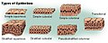
Classification

In general, tissues are classified by the morphology of their cells, and the number of layers they are composed of.[2][4][6] Epithelial tissue that is only one cell thick is known as simple epithelium.[7] If it is two or more cells thick, it is known as stratified epithelium.[8] However, when taller simple epithelial cells (see columnar, below) are viewed in cross section with several nuclei appearing at different heights, they can be confused with stratified epithelia. This kind of epithelium is therefore described as "pseudostratified" epithelium.[9]
There are three principal morphologies associated with epithelial cells. Squamous epithelium has cells that are wider than they are tall (flat and scale-like). Cuboidal epithelium has cells whose height and width are approximately the same (cube shaped). Columnar epithelium has cells taller than they are wide (column-shaped). In addition, the morphology of the cells in transitional epithelium may vary from squamous to cuboidal, depending on the amount of tension on the epithelium.[10]
Simple epithelium
Simple epithelium is one cell thick; that is, every cell is in direct contact with the underlying basement membrane. In general, it is found where absorption and filtration occur. The thinness of the epithelial barrier facilitates these processes.[4]
In general, simple epithelial tissues are classified by the shape of their cells. The four major classes of simple epithelium are: (1) simple squamous; (2) simple cuboidal; (3) simple columnar; (4) pseudostratified.[4]
Simple squamous epithelium is found lining areas where passive diffusion of gases occur, including the walls of capillaries, the linings of the alveoli of the lungs, and the linings of the pericardial, pleural, and peritoneal cavities.
Functions
The primary functions of epithelial tissues are: (1) to protect the tissues that lie beneath it from radiation, desiccation, toxins, invasion by pathogens, and physical trauma; (2) the regulation and exchange of chemicals between the underlying tissues and a body cavity; (3) the secretion of hormones into the blood vascular system, and/or the secretion of sweat, mucus, enzymes, and other products that are delivered by ducts glandular epithelium;[11] (4) to provide sensation.
Secretory epithelia


As stated above, secretion is one major function of epithelial cells. Glands are formed from the invagination / infolding of epithelial cells and subsequent growth in the underlying connective tissue. There are two major classifications of glands: endocrine glands and exocrine glands. Endocrine glands secrete their product into the extracellular space where it is rapidly taken up by the blood vascular system. The exocrine glands secrete their products into a duct that then delivers the product to the lumen of an organ or onto the free surface of the epithelium.
In arthropods, the integument, or external "skin", consists of a single layer of epithelial ectoderm from which arises the cuticle,[12] an outer covering of chitin the rigidity of which varies as per its chemical composition.
Sensing the extracellular environment
"Some epithelial cells are ciliated, and they commonly exist as a sheet of polarised cells forming a tube or tubule with cilia projecting into the lumen." Primary cilia on epithelial cells provide chemosensation, thermosensation, and mechanosensation of the extracellular environment by playing "a sensory role mediating specific signalling cues, including soluble factors in the external cell environment, a secretory role in which a soluble protein is released to have an effect downstream of the fluid flow, and mediation of fluid flow if the cilia are motile."[13]
Embryological development
In general, there are epithelial tissues deriving from all of the embryological germ layers:[citation needed]
- from ectoderm (e.g., the epidermis);
- from endoderm (e.g., the lining of the gastrointestinal tract);
- from mesoderm (e.g., the inner linings of body cavities).
However, it is important to note that pathologists do not consider endothelium and mesothelium (both derived from mesoderm) to be true epithelium. This is because such tissues present very different pathology. For that reason, pathologists label cancers in endothelium and mesothelium sarcomas, whereas true epithelial cancers are called carcinomas. Also, the filaments that support these mesoderm-derived tissues are very distinct. Outside of the field of pathology, it is, in general, accepted that the epithelium arises from all three germ layers.[citation needed]
Growing in culture
When growing epithelium in culture, one can determine whether or not a particular cell is epithelial by examining its morphological characteristics. Epithelial cells tend to cluster together, and have a "characteristic tight pavementlike appearance". But this is not always the case, such as when the cells are derived from a tumor. In these cases, it is often necessary to use certain biochemical markers to make a positive identification. The intermediate filament proteins in the cytokeratin group are almost exclusively found in epithelial cells, and so are often used for this purpose.[14]
Location
Epithelium lines both the outside (skin) and the inside cavities and lumen of bodies. The outermost layer of our skin is composed of dead stratified squamous, keratinized epithelial cells.[citation needed]
Tissues that line the inside of the mouth, the esophagus and part of the rectum are composed of nonkeratinized stratified squamous epithelium. Other surfaces that separate body cavities from the outside environment are lined by simple squamous, columnar, or pseudostratified epithelial cells. Other epithelial cells line the insides of the lungs, the gastrointestinal tract, the reproductive and urinary tracts, and make up the exocrine and endocrine glands. The outer surface of the cornea is covered with fast-growing, easily regenerated epithelial cells. Endothelium (the inner lining of blood vessels, the heart, and lymphatic vessels) is a specialized form of epithelium. Another type, mesothelium, forms the walls of the pericardium, pleurae, and peritoneum.[citation needed]
 |
| Part of a series of lists about |
| Human anatomy |
|---|
| This article is part of a series on |
| Epithelia |
|---|
| Squamous epithelial cell |
| Columnar epithelial cell |
| Cuboidal epithelial cell |
| Specialised epithelia |
|
| Other |
This table lists the epithelia of different organs of the human body
| System | Tissue | Epithelium | Subtype |
|---|---|---|---|
| circulatory | blood vessels | Simple squamous | endothelium |
| digestive | ducts of submandibular glands | simple columnar | - |
| digestive | attached gingiva | Stratified squamous, keratinized | - |
| digestive | dorsum of tongue | Stratified squamous, keratinized | - |
| digestive | hard palate | Stratified squamous, keratinized | - |
| digestive | oesophagus | Stratified squamous, non-keratinized | - |
| digestive | stomach | Simple columnar, non-ciliated | gastric epithelium |
| digestive | small intestine | Simple columnar, non-ciliated | intestinal epithelium |
| digestive | large intestine | Simple columnar, non-ciliated | intestinal epithelium |
| digestive | rectum | Simple columnar, non-ciliated | - |
| digestive | anus | Stratified squamous, non-keratinized superior to Hilton's white line Stratified squamous, keratinized inferior to Hilton's white line |
- |
| digestive | gallbladder | Simple columnar, non-ciliated | - |
| endocrine | thyroid follicles | Simple cuboidal | - |
| nervous | ependyma | Simple cuboidal | - |
| lymphatic | lymph vessel | Simple squamous | endothelium |
| integumentary | skin - dead superficial layer | Stratified squamous, keratinized | - |
| integumentary | sweat gland ducts | Stratified cuboidal | - |
| integumentary | mesothelium of body cavities | Simple squamous | mesothelium |
| reproductive - female | ovaries | Simple cuboidal | germinal epithelium (female) |
| reproductive - female | fallopian tubes | Simple columnar, ciliated | - |
| reproductive - female | endometrium (uterus) | Simple columnar, ciliated | - |
| reproductive - female | cervix (endocervix) | Simple columnar | - |
| reproductive - female | cervix (ectocervix) | Stratified squamous, non-keratinized | - |
| reproductive - female | vaginal epithelium | Stratified squamous, non-keratinized | - |
| reproductive - female | labia majora | Stratified squamous, keratinized | - |
| reproductive - male | tubuli recti | Simple cuboidal | germinal epithelium (male) |
| reproductive - male | rete testis | Simple cuboidal | - |
| reproductive - male | efferent ducts | Pseudostratified columnar | - |
| reproductive - male | epididymis | Pseudostratified columnar, with stereocilia | - |
| reproductive - male | vas deferens | Pseudostratified columnar | - |
| reproductive - male | ejaculatory duct | Simple columnar | - |
| reproductive - male (gland) | bulbourethral glands | Simple columnar | - |
| reproductive - male (gland) | seminal vesicle | Pseudostratified columnar | - |
| respiratory | oropharynx | Stratified squamous, non-keratinized | - |
| respiratory | larynx | Pseudostratified columnar, ciliated | respiratory epithelium |
| respiratory | larynx - true vocal cords | Stratified squamous, non-keratinized | - |
| respiratory | trachea | Pseudostratified columnar, ciliated | respiratory epithelium |
| respiratory | bronchi | Pseudostratified columnar, ciliated | |
| respiratory | terminal bronchioles | Simple cuboidal, ciliated | |
| respiratory | respiratory bronchioles | Simple cuboidal, ciliated | - |
| respiratory | alveoli | Simple squamous | |
| sensory | cornea | Stratified squamous, non-keratinized | corneal epithelium |
| sensory | nose | Pseudostratified columnar | olfactory epithelium |
| urinary | kidney - proximal convoluted tubule | Simple cuboidal, with microvilli | - |
| urinary | kidney - ascending thin limb | Simple squamous | - |
| urinary | kidney - distal convoluted tubule | Simple cuboidal, without microvilli | - |
| urinary | kidney - collecting duct | Simple cuboidal | - |
| urinary | kidney - Bowman's capsule | Simple squamous | - |
| urinary | kidney - Loop of Henle | Simple squamous | - |
| urinary | kidney - descending thin limb | Simple squamous | - |
| urinary | kidney - descending thick limb | simple cuboidal | - |
| urinary | renal pelvis | Transitional | urothelium |
| urinary | ureter | Transitional | urothelium |
| urinary | urinary bladder | Transitional | urothelium |
| urinary | prostatic urethra | Transitional | urothelium |
| urinary | membranous urethra | Pseudostratified columnar, non-ciliated | - |
| urinary | penile urethra | Pseudostratified columnar, non-ciliated | - |
| urinary | urinary meatus | Stratified squamous | - |
Additional images
-
 Squamous epithelium is one of several types of epithelia.
 -
 Squamous Epithelium 100x
 -
 Human cheek cells (Nonkeratinized stratified squamous epithelium) 500x


See also
References
Notes
- ^ Epithelium at Wiktionary
- ^ a b c Dellmann's textbook of veterinary histology. Wiley-Blackwell. 2006. p. 18. ISBN 978-0-7817-4148-4.
{{cite book}}: Unknown parameter|editors=ignored (|editor=suggested) (help) - ^ a b Freshney, 2002: p. 3
- ^ a b c d Marieb, Elaine M. (1995). Human Anatomy and Physiology (3rd ed.). Benjamin/Cummings. pp. 103–104. ISBN 0-8053-4281-8.
- ^ McConnell, Thomas H. (2006). The nature of disease: pathology for the health professions. Lippincott Williams & Wilkins. p. 55. ISBN 978-0-7817-5317-3.
- ^ Platzer, Werner (2008). Color atlas of human anatomy: Locomotor system. Thieme. p. 8. ISBN 978-3-13-533306-9.
- ^ van Lommel, 2002: p. 94
- ^ van Lommel, 2002: p. 97
- ^ Permar's oral embryology and microscopic anatomy: a textbook for students in dental hygiene. Lippincott Williams & Wilkins. 2000. p. 9. ISBN 978-0-683-30644-6.
{{cite book}}: Unknown parameter|editors=ignored (|editor=suggested) (help) - ^ Pratt, Rebecca. "Epithelial Cells". AnatomyOne. Amirsys, Inc. Retrieved 2012-09-28.
- ^ van Lommel, 2002: p. 91
- ^ Kristensen, Niels P.; Georges, Chauvin (1 December 2003). "Integument". Lepidoptera, Moths and Butterflies: Morphology, Physiology, and Development : Teilband. Walter de Gruyter. p. 484. ISBN 978-3-11-016210-3. Retrieved 10 January 2013.
- ^ Adams, M.; Smith, U.M.; Logan, C.V.; Johnson, C.A. (2008). "Recent advances in the molecular pathology, cell biology and genetics of ciliopathies". Journal of Medical Genetics. 45 (5): 257–267. doi:10.1136/jmg.2007.054999. PMID 18178628.
{{cite journal}}: CS1 maint: multiple names: authors list (link) - ^ Freshney, 2002: p. 9
Bibliography
- Freshney, R.I. (2002). "Introduction". Culture of epithelial cells. John Wiley & Sons. ISBN 978-0-471-40121-6.
{{cite book}}: Unknown parameter|editors=ignored (|editor=suggested) (help) - van Lommel, Alfons T.L. (2002). From cells to organs: a histology textbook and atlas. Springer. ISBN 978-1-4020-7257-4.
Further reading
- Green H (September 2008). "The birth of therapy with cultured cells". BioEssays. 30 (9): 897–903. doi:10.1002/bies.20797. PMID 18693268.
- Basement membranes: cell and molecular biology. Gulf Professional Publishing. 2005. ISBN 978-0-12-153356-4.
{{cite book}}: Unknown parameter|editors=ignored (|editor=suggested) (help) - Nagpal R, Patel A, Gibson MC (March 2008). "Epithelial topology". BioEssays. 30 (3): 260–6. doi:10.1002/bies.20722. PMID 18293365.
{{cite journal}}: CS1 maint: multiple names: authors list (link) - Yamaguchi Y, Brenner M, Hearing VJ (September 2007). "The regulation of skin pigmentation" (Review). J. Biol. Chem. 282 (38): 27557–61. doi:10.1074/jbc.R700026200. PMID 17635904.
{{cite journal}}: CS1 maint: multiple names: authors list (link) CS1 maint: unflagged free DOI (link)
| Animals | |
|---|---|
| Plants | |