
Brodmann area: Difference between revisions
→External links: Removed broken links Tags: Mobile edit Mobile web edit |
Rescuing 1 sources and tagging 1 as dead. #IABot (v1.2.7) |
||
| Line 153: | Line 153: | ||
* [http://www.brodmannarea.info/index2.htm] - Brodmann Areas, their functions, and the lateralization of functions across hemispheres |
* [http://www.brodmannarea.info/index2.htm] - Brodmann Areas, their functions, and the lateralization of functions across hemispheres |
||
* [http://spot.colorado.edu/~dubin/talks/brodmann/brodmann.html Brodmann], Mark Dubin pages on Brodmann areas. |
* [http://spot.colorado.edu/~dubin/talks/brodmann/brodmann.html Brodmann], Mark Dubin pages on Brodmann areas. |
||
* [http://braininfo.rprc.washington.edu/indexotheratlas.aspx?othersiteID=1045244870 Brodmann areas] Brodmann areas of cortex involved in language. |
* [https://web.archive.org/web/20120111073154/http://braininfo.rprc.washington.edu:80/indexotheratlas.aspx?othersiteID=1045244870 Brodmann areas] Brodmann areas of cortex involved in language. |
||
* [http://braininfo.rprc.washington.edu/ShowItHier.aspx?questID=1032 Illustrations] More Illustrations. |
* [http://braininfo.rprc.washington.edu/ShowItHier.aspx?questID=1032 Illustrations]{{dead link|date=November 2016 |bot=InternetArchiveBot |fix-attempted=yes }} More Illustrations. |
||
{{Brodmann area}} |
{{Brodmann area}} |
||
Revision as of 05:01, 9 November 2016

A Brodmann area is a region of the cerebral cortex, in the human or other primate brain, defined by its cytoarchitecture, or histological structure and organization of cells.
History

Brodmann areas were originally defined and numbered by the German anatomist Korbinian Brodmann based on the cytoarchitectural organization of neurons he observed in the cerebral cortex using the Nissl method of cell staining. Brodmann published his maps of cortical areas in humans, monkeys, and other species in 1909,[1] along with many other findings and observations regarding the general cell types and laminar organization of the mammalian cortex. The same Brodmann area number in different species does not necessarily indicate homologous areas.[2] A similar, but more detailed cortical map was published by Constantin von Economo and Georg N. Koskinas in 1925.[3]
Present importance
Brodmann areas have been discussed, debated, refined, and renamed exhaustively for nearly a century and remain the most widely known and frequently cited cytoarchitectural organization of the human cortex.
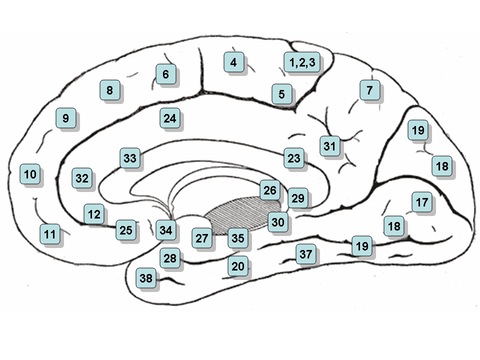
Many of the areas Brodmann defined based solely on their neuronal organization have since been correlated closely to diverse cortical functions. For example, Brodmann areas 1, 2 and 3 are the primary somatosensory cortex; area 4 is the primary motor cortex; area 17 is the primary visual cortex; and areas 41 and 42 correspond closely to primary auditory cortex. Higher order functions of the association cortical areas are also consistently localized to the same Brodmann areas by neurophysiological, functional imaging, and other methods (e.g., the consistent localization of Broca's speech and language area to the left Brodmann areas 44 and 45). However, functional imaging can only identify the approximate localization of brain activations in terms of Brodmann areas since their actual boundaries in any individual brain requires its histological examination.
Brodmann areas for humans and other primates
- Areas 3, 1 & 2 – Primary Somatosensory Cortex (frequently referred to as Areas 3, 1, 2 by convention)
- Area 4 – Primary Motor Cortex
- Area 5 – Somatosensory Association Cortex
- Area 6 – Premotor cortex and Supplementary Motor Cortex (Secondary Motor Cortex) (Supplementary motor area)
- Area 7 – Visuo-Motor Coordination
- Area 8 – Includes Frontal eye fields
- Area 9 – Dorsolateral prefrontal cortex
- Area 10 – Anterior prefrontal cortex (most rostral part of superior and middle frontal gyri)
- Area 11 – Orbitofrontal area (orbital and rectus gyri, plus part of the rostral part of the superior frontal gyrus)
- Area 12 – Orbitofrontal area (used to be part of BA11, refers to the area between the superior frontal gyrus and the inferior rostral sulcus)
- Area 13 and Area 14* – Insular cortex
- Area 15* – Anterior Temporal lobe
- Area 16 – Insular cortex
- Area 17 – Primary visual cortex (V1)
- Area 18 – Secondary visual cortex (V2)
- Area 19 – Associative visual cortex (V3,V4,V5)
- Area 20 – Inferior temporal gyrus
- Area 21 – Middle temporal gyrus
- Area 22 – Superior temporal gyrus, of which the caudal part is usually considered to contain the Wernicke's area
- Area 23 – Ventral posterior cingulate cortex
- Area 24 – Ventral anterior cingulate cortex.
- Area 25 – Subgenual area (part of the Ventromedial prefrontal cortex)[4]
- Area 26 – Ectosplenial portion of the retrosplenial region of the cerebral cortex
- Area 27 – Piriform cortex
- Area 28 – Ventral entorhinal cortex
- Area 29 – Retrosplenial cingulate cortex
- Area 30 – Part of cingulate cortex
- Area 31 – Dorsal Posterior cingulate cortex
- Area 32 – Dorsal anterior cingulate cortex
- Area 33 – Part of anterior cingulate cortex
- Area 34 – Dorsal entorhinal cortex (on the Parahippocampal gyrus)
- Area 35 – Perirhinal cortex (in the rhinal sulcus)
- Area 36 – Ectorhinal area, now part of the perirhinal cortex (in the rhinal sulcus)
- Area 37 – Fusiform gyrus
- Area 38 – Temporopolar area (most rostral part of the superior and middle temporal gyri)
- Area 39 – Angular gyrus, considered by some to be part of Wernicke's area
- Area 40 – Supramarginal gyrus considered by some to be part of Wernicke's area
- Areas 41 and 42 – Auditory cortex
- Area 43 – Primary gustatory cortex
- Area 44 – Pars opercularis, part of the inferior frontal gyrus and part of Broca's area
- Area 45 – Pars triangularis, part of the inferior frontal gyrus and part of Broca's area
- Area 46 – Dorsolateral prefrontal cortex
- Area 47 – Pars orbitalis, part of the inferior frontal gyrus
- Area 48 – Retrosubicular area (a small part of the medial surface of the temporal lobe)
- Area 49 – Parasubicular area in a rodent
- Area 52 – Parainsular area (at the junction of the temporal lobe and the insula)
(*) Area only found in non-human primates.
Some of the original Brodmann areas have been subdivided further, e.g., "23a" and "23b".[5]
Clickable map: lateral surface
- Note: the lateral view, or side view, of the brain is denoted the 'lateral surface'

Clickable map: medial surface
- Note: the view of the section between the right and left hemispheres of the brain is denoted the 'medial surface'
Criticism
When von Bonin and Bailey constructed a brain map for the macaque monkey they found the description of Brodmann inadequate and wrote: "Brodmann (1907), it is true, prepared a map of the human brain which has been widely reproduced, but, unfortunately, the data on which it was based was never published"[6] They instead used the cytoarchitechtonic scheme of Constantin von Economo and Georg N. Koskinas published in 1925[3] which had the "only acceptable detailed description of the human cortex".
See also
References
- ^ Brodmann K (1909). "Vergleichende Lokalisationslehre der Grosshirnrinde" (in German). Leipzig: Johann Ambrosius Barth.
{{cite journal}}: Cite journal requires|journal=(help)[page needed] - ^ Garey LJ. (2006). Brodmann's Localisation in the Cerebral Cortex. New York: Springer. ISBN 978-0387-26917-7.[page needed]
- ^ a b Economo, C.; Koskinas, G.N. (1925). "Die Cytoarchitektonik der Hirnrinde des erwachsenen Menschen" (in German). Wien & Berlin: Springer.
{{cite journal}}: Cite journal requires|journal=(help)[page needed] - ^ Fales CL, Barch DM, Rundle MM, Mintun MA, Snyder AZ, Cohen JD, Mathews J, Sheline YI (February 2008). "Altered emotional interference processing in affective and cognitive-control brain circuitry in major depression". Biol. Psychiatry. 63 (4): 377–84. doi:10.1016/j.biopsych.2007.06.012. PMC 2268639. PMID 17719567.
- ^ Brent A. Vogt; Deepak N. Pandya; Douglas L. Rosene (August 1987). "Cingulate Cortex of the Rhesus Monkey: I. Cytoarchitecture and Thalamic Afferents". The Journal of Comparative Neurology. 262 (2): 256–270. doi:10.1002/cne.902620207. PMID 3624554.
- ^ Gerhardt von Bonin; Percival Bailey (1925). The Neocortex of Macaca Mulatta (PDF). Urbana, Illinois: The University of Illinois Press.
{{cite book}}: Unknown parameter|last-author-amp=ignored (|name-list-style=suggested) (help)
External links
- [1] - Brodmann Areas, their functions, and the lateralization of functions across hemispheres
- Brodmann, Mark Dubin pages on Brodmann areas.
- Brodmann areas Brodmann areas of cortex involved in language.
- Illustrations[permanent dead link] More Illustrations.