
Lacrimal bone
| Lacrimal bone | |
|---|---|
 Lacrimal bone visible near center. | |
 Orbital bones. Lacrimal bone shown in green. | |
| Details | |
| Identifiers | |
| Latin | os lacrimale |
| TA98 | A02.1.09.001 |
| TA2 | 744 |
| FMA | 52741 |
| Anatomical terms of bone | |
The lacrimal bone, the smafllest and most fragile bone of the face, is situated at the front part of the medial wall of the orbit. It has two surfaces and four borders.
Surfaces
Lateral or orbital surface
The lateral or orbital surface is divided by a vertical ridge, the posterior lacrimal crest, into two parts.
In front of this crest is a longitudinal groove, the lacrimal sulcus (sulcus lacrimalis), the inner margin of which unites with the frontal process of the maxilla, and the lacrimal fossa is thus completed. The upper part of this fossa lodges the lacrimal sac, the lower part, the nasolacrimal duct.
The portion behind the crest is smooth, and forms part of the medial wall of the orbit.
The crest, with a part of the orbital surface immediately behind it, gives origin to the lacrimal part of the Orbicularis oculi and ends below in a small, hook-like projection, the lacrimal hamulus, which articulates with the lacrimal tubercle of the maxilla, and completes the upper orifice of the nasolacrimal canal; the hamulus sometimes exists as a separate piece, and is then called the lesser lacrimal bone.
Medial or nasal surface
The medial or nasal surface presents a longitudinal furrow, corresponding to the crest on the lateral surface.
The area in front of this furrow forms part of the middle meatus of the nose; that behind it articulates with the ethmoid, and completes some of the anterior ethmoidal cells.
Borders
Of the four borders:
- the anterior articulates with the frontal process of the maxilla;
- the posterior with the lamina papyracea of the ethmoid;
- the superior with the frontal bone.
- The inferior is divided by the lower edge of the posterior lacrimal crest into two parts:
- the posterior part articulates with the orbital plate of the maxilla;
- the anterior is prolonged downward as the descending process, which articulates with the lacrimal process of the inferior nasal concha, and assists in forming the canal for the nasolacrimal duct.
Ossification
The lacrimal is ossified from a single center, which appears about the twelfth week in the membrane covering the cartilaginous nasal capsule.
Articulations
The lacrimal articulates with four bones: two of the cranium, the frontal and ethmoid, and two of the face, the maxilla and the inferior nasal concha.
In other animals
In early lobe-finned fishes and ancestral tetrapods, the lacrimal bone is a relatively large and robust bone, running from the orbit to the nostrils. It forms part of the side of the face, between the nasal bones and the maxilla. In primitive forms, it is often accompanied by a much smaller septomaxilla bone, lying immediately behind the nasal opening, but this is lost in most modern species. The lacrimal bone is often smaller in living vertebrates, and is no longer always directly associated with the nasal opening, although it retains its connection with the orbit. The bone is entirely absent in living amphibians, as well as some reptilian species.[1]
Additional images
-
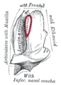 Left lacrimal bone. Orbital surface. Enlarged
 -
 Side view of the skull.
 -
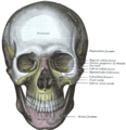 The skull from the front.
 -
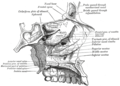
 Medial wall of left orbit.
 -
 Roof, floor, and lateral wall of left nasal cavity.


See also
External links
- Template:EMedicineDictionary
- Atlas image: eye_5 at the University of Michigan Health System
- Atlas image: rsa2p4 at the University of Michigan Health System
- Template:RocheLexicon
- Diagram at upstate.edu
References
- ^ Romer, Alfred Sherwood; Parsons, Thomas S. (1977). The Vertebrate Body. Philadelphia, PA: Holt-Saunders International. pp. 217–241. ISBN 0-03-910284-X.