Thrombospondin 1
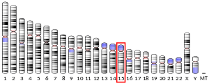
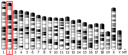
Thrombospondin 1, abbreviated as THBS1, is a protein that in humans is encoded by the THBS1 gene.[5][6]
Thrombospondin 1 is a subunit of a disulfide-linked homotrimeric protein. This protein is an adhesive glycoprotein that mediates cell-to-cell and cell-to-matrix interactions. This protein can bind to fibrinogen, fibronectin, laminin, collagens types V and VII and integrins alpha-V/beta-1. This protein has been shown to play roles in platelet aggregation, angiogenesis, and tumorigenesis.[7][8]
Function
The thrombospondin-1 protein is a member of the thrombospondin family. It is a multi-domain matrix glycoprotein that has been shown to be a natural inhibitor of neovascularization and tumorigenesis in healthy tissue. Both positive and negative modulation of endothelial cell adhesion, motility, and growth have been attributed to TSP1. This should not be surprising considering that TSP1 interacts with at least 12 cell adhesion receptors, including CD36, αv integrins, β1 integrins, syndecan, and integrin-associated protein (IAP or CD47). It also interacts with numerous proteases involved in angiogenesis, including plasminogen, urokinase, matrix metalloproteinase, thrombin, cathepsin, and elastase.
Thrombospondin-1 binds to the reelin receptors, ApoER2 and VLDLR, thereby affecting neuronal migration in the rostral migratory stream.[9]
The various functions of the TSRs have been attributed to several recognition motifs. Characterization of these motifs has led to the use of recombinant proteins that contain these motifs; these recombinant proteins are deemed useful in cancer therapy. The TSP-1 3TSR (a recombinant version of the THBS1 antiangiogenic domain containing all three thrombosopondin-1 type 1 repeats) can activate transforming growth factor beta 1 (TGFβ1) and inhibit endothelial cell migration, angiogenesis, and tumor growth.[10]
Structure
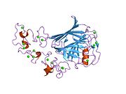
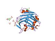
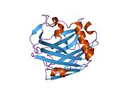
Thrombospondin's activity has been mapped to several domains, in particular the amino-terminal heparin-binding domain, the procollagen domain, the properdin-like type I repeats, and the globular carboxy-terminal domain. The protein also contains type II repeats with epidermal growth factor-like homology and type III repeats that contain an RGD sequence.[11]
N-terminus
The N-terminal heparin-binding domain of TSP1, when isolated as a 25kDa fragment, has been shown to be a potent inducer of cell migration at high concentrations. However, when the heparin-binding domain of TSP1 is cleaved, the remaining anti-angiogenic domains have been shown to have decreased anti-angiogenic activity at low concentrations where increased endothelial cell (EC) migration occurs. This may be explained in part by the ability of the heparin-binding domain to mediate attachment of TSP1 to cells, allowing the other domains to exert their effects. The separate roles that the heparin-binding region of TSP1 plays at high versus low concentrations may be in part responsible for regulating the two-faced nature of TSP1 and giving it a reputation of being both a positive and negative regulator of angiogenesis.[12]
Procollagen domain
Both the procollagen domain and the type I repeats of TSP1 have been shown to inhibit neovascularization and EC migration. However, it is unlikely that the mechanisms of action of these fragments are the same. The type I repeats of TSP1 are capable of inhibiting EC migration in a Boyden chamber assay after a 3-4 hour exposure, whereas a 36- to 48-hour exposure period is necessary for inhibition of EC migration with the procollagen domain.[12] Whereas the chorioallantoic membrane (CAM) assay shows the type I repeats of TSP1 to be antiangiogenic, it also shows that the procollagen sequence lacks anti-angiogenic activity. This may be in part because the animo-terminal end of TSP1 differs more than the carboxy-terminal end across species, but may also suggest different mechanisms of action.[13]
TSP1 contains three type I repeats, only the second two of which have been found to inhibit angiogenesis. The type I repeat motif is more effective than the entire protein at inhibiting angiogenesis and contains not one but two regions of activity. The amino-terminal end contains a tryptophan-rich motif that blocks fibroblast growth factor (FGF-2 or bFGF) driven angiogenesis. This region has also been found to prevent FGF-2 binding ECs, suggesting that its mechanism of action may be to sequester FGF-2. The second region of activity, the CD36 binding region of TSP1, can be found on the carboxy-terminal half of the type I repeats.[13] It has been suggested that activating the CD36 receptor causes an increase in ECs sensitivity to apoptotic signals.[14][15] Type I repeats have also been shown to bind to heparin, fibronectin, TGF-β, and others, potentially antagonizing the effects of these molecules on ECs.[16] However, CD36 is generally considered to be the dominant inhibitory signaling receptor for TSP1, and EC expression of CD36 is restricted to microvascular ECs.
Soluble type I repeats have been shown to decrease EC numbers by inhibiting proliferation and promoting apoptosis. Attachment of endothelial cells to fibronectin partially reverses this phenomenon. However this domain is not without a two-faced nature of its own. Bound protein fragments of the type I repeats have been shown to serve as attachment factors for both ECs and melanoma cells.[17]
C-terminus
The carboxy-terminal domain of TSP1 is believed to mediate cellular attachment and has been found to bind to another important receptor for TSP1, IAP (or CD47).[18] This receptor is considered necessary for nitric oxide-stimulated TSP1-mediated vascular cell responses and cGMP signaling.[19] Various domains of and receptors for TSP1 have been shown to have pro-adhesive and chemotactic activities for cancer cells, suggesting that this molecule may have a direct effect on cancer cell biology independent of its anti-angiogenic properties.[20][21]
Cancer treatment
One study has suggested that, by blocking TSP1 from binding to its cell surface receptor (CD47) normal tissue confers high resistance to cancer radiation therapy and assists in tumor death.[22]
Interactions
Thrombospondin 1 has been shown to interact with:
References
- ^ a b c GRCh38: Ensembl release 89: ENSG00000137801 – Ensembl, May 2017
- ^ a b c GRCm38: Ensembl release 89: ENSMUSG00000040152 – Ensembl, May 2017
- ^ "Human PubMed Reference:". National Center for Biotechnology Information, U.S. National Library of Medicine.
- ^ "Mouse PubMed Reference:". National Center for Biotechnology Information, U.S. National Library of Medicine.
- ^ Wolf FW, Eddy RL, Shows TB, Dixit VM (Apr 1990). "Structure and chromosomal localization of the human thrombospondin gene" (PDF). Genomics. 6 (4): 685–91. doi:10.1016/0888-7543(90)90505-O. hdl:2027.42/28657. PMID 2341158.
- ^ Jaffe E, Bornstein P, Disteche CM (May 1990). "Mapping of the thrombospondin gene to human chromosome 15 and mouse chromosome 2 by in situ hybridization". Genomics. 7 (1): 123–6. doi:10.1016/0888-7543(90)90528-3. PMID 2335352.
- ^ "Entrez Gene: THBS1 thrombospondin 1".
- ^ Atanasova, VS; Russell, RJ; Webster, TG; Cao, Q; Agarwal, P; Lim, YZ; Krishnan, S; Fuentes, I; Guttmann-Gruber, C; McGrath, JA; Salas-Alanis, JC; Fertala, A; South, AP (July 2019). "Thrombospondin-1 Is a Major Activator of TGF-β Signaling in Recessive Dystrophic Epidermolysis Bullosa Fibroblasts". The Journal of Investigative Dermatology. 139 (7): 1497–1505.e5. doi:10.1016/j.jid.2019.01.011. PMID 30684555.
- ^ Blake SM, Strasser V, Andrade N, Duit S, Hofbauer R, Schneider WJ, Nimpf J (Nov 2008). "Thrombospondin-1 binds to ApoER2 and VLDL receptor and functions in postnatal neuronal migration". The EMBO Journal. 27 (22): 3069–80. doi:10.1038/emboj.2008.223. PMC 2585172. PMID 18946489.
- ^ Lopez-Dee ZP, Chittur SV, Patel B, Stanton R, Wakeley M, Lippert B, Menaker A, Eiche B, Terry R, Gutierrez LS (2012). "Thrombospondin-1 type 1 repeats in a model of inflammatory bowel disease: transcript profile and therapeutic effects". PLOS ONE. 7 (4): e34590. Bibcode:2012PLoSO...734590L. doi:10.1371/journal.pone.0034590. PMC 3318003. PMID 22509329.
{{cite journal}}: CS1 maint: unflagged free DOI (link) - ^ Forslöw A, Liu Z, Sundqvist KG (Jan 2007). "Receptor communication within the lymphocyte plasma membrane: a role for the thrombospondin family of matricellular proteins". Cellular and Molecular Life Sciences. 64 (1): 66–76. doi:10.1007/s00018-006-6255-8. PMID 17160353.
- ^ a b Tolsma SS, Volpert OV, Good DJ, Frazier WA, Polverini PJ, Bouck N (Jul 1993). "Peptides derived from two separate domains of the matrix protein thrombospondin-1 have anti-angiogenic activity". The Journal of Cell Biology. 122 (2): 497–511. doi:10.1083/jcb.122.2.497. PMC 2119646. PMID 7686555.
- ^ a b Iruela-Arispe ML, Lombardo M, Krutzsch HC, Lawler J, Roberts DD (Sep 1999). "Inhibition of angiogenesis by thrombospondin-1 is mediated by 2 independent regions within the type 1 repeats". Circulation. 100 (13): 1423–31. doi:10.1161/01.cir.100.13.1423. PMID 10500044.
- ^ Guo N, Krutzsch HC, Inman JK, Roberts DD (May 1997). "Thrombospondin 1 and type I repeat peptides of thrombospondin 1 specifically induce apoptosis of endothelial cells". Cancer Research. 57 (9): 1735–42. PMID 9135017.
- ^ Sid B, Sartelet H, Bellon G, El Btaouri H, Rath G, Delorme N, Haye B, Martiny L (Mar 2004). "Thrombospondin 1: a multifunctional protein implicated in the regulation of tumor growth". Critical Reviews in Oncology/Hematology. 49 (3): 245–58. doi:10.1016/j.critrevonc.2003.09.009. PMID 15036264.
- ^ Guo N, Zabrenetzky VS, Chandrasekaran L, Sipes JM, Lawler J, Krutzsch HC, Roberts DD (Jul 1998). "Differential roles of protein kinase C and pertussis toxin-sensitive G-binding proteins in modulation of melanoma cell proliferation and motility by thrombospondin 1". Cancer Research. 58 (14): 3154–62. PMID 9679984.
- ^ Prater CA, Plotkin J, Jaye D, Frazier WA (Mar 1991). "The properdin-like type I repeats of human thrombospondin contain a cell attachment site". The Journal of Cell Biology. 112 (5): 1031–40. doi:10.1083/jcb.112.5.1031. PMC 2288870. PMID 1999454.
- ^ Kosfeld MD, Frazier WA (Aug 1992). "Identification of active peptide sequences in the carboxyl-terminal cell binding domain of human thrombospondin-1". The Journal of Biological Chemistry. 267 (23): 16230–6. PMID 1644809.
- ^ Isenberg JS, Ridnour LA, Dimitry J, Frazier WA, Wink DA, Roberts DD (Sep 2006). "CD47 is necessary for inhibition of nitric oxide-stimulated vascular cell responses by thrombospondin-1". The Journal of Biological Chemistry. 281 (36): 26069–80. doi:10.1074/jbc.M605040200. PMID 16835222.
- ^ Chandrasekaran S, Guo NH, Rodrigues RG, Kaiser J, Roberts DD (Apr 1999). "Pro-adhesive and chemotactic activities of thrombospondin-1 for breast carcinoma cells are mediated by alpha3beta1 integrin and regulated by insulin-like growth factor-1 and CD98". The Journal of Biological Chemistry. 274 (16): 11408–16. doi:10.1074/jbc.274.16.11408. PMID 10196234.
- ^ Taraboletti G, Roberts DD, Liotta LA (Nov 1987). "Thrombospondin-induced tumor cell migration: haptotaxis and chemotaxis are mediated by different molecular domains". The Journal of Cell Biology. 105 (5): 2409–15. doi:10.1083/jcb.105.5.2409. PMC 2114831. PMID 3680388.
- ^ Maxhimer JB, Soto-Pantoja DR, Ridnour LA, Shih HB, Degraff WG, Tsokos M, Wink DA, Isenberg JS, Roberts DD (Oct 2009). "Radioprotection in normal tissue and delayed tumor growth by blockade of CD47 signaling". Science Translational Medicine. 1 (3): 3ra7. doi:10.1126/scitranslmed.3000139. PMC 2811586. PMID 20161613.
{{cite journal}}: Unknown parameter|lay-url=ignored (help); Unknown parameter|laysource=ignored (help) - ^ Wang S, Herndon ME, Ranganathan S, Godyna S, Lawler J, Argraves WS, Liau G (Mar 2004). "Internalization but not binding of thrombospondin-1 to low density lipoprotein receptor-related protein-1 requires heparan sulfate proteoglycans". Journal of Cellular Biochemistry. 91 (4): 766–76. doi:10.1002/jcb.10781. PMID 14991768.
- ^ Mikhailenko I, Krylov D, Argraves KM, Roberts DD, Liau G, Strickland DK (Mar 1997). "Cellular internalization and degradation of thrombospondin-1 is mediated by the amino-terminal heparin binding domain (HBD). High affinity interaction of dimeric HBD with the low density lipoprotein receptor-related protein". The Journal of Biological Chemistry. 272 (10): 6784–91. doi:10.1074/jbc.272.10.6784. PMID 9045712.
- ^ Godyna S, Liau G, Popa I, Stefansson S, Argraves WS (Jun 1995). "Identification of the low density lipoprotein receptor-related protein (LRP) as an endocytic receptor for thrombospondin-1". The Journal of Cell Biology. 129 (5): 1403–10. doi:10.1083/jcb.129.5.1403. PMC 2120467. PMID 7775583.
- ^ Bein K, Simons M (Oct 2000). "Thrombospondin type 1 repeats interact with matrix metalloproteinase 2. Regulation of metalloproteinase activity". The Journal of Biological Chemistry. 275 (41): 32167–73. doi:10.1074/jbc.M003834200. PMID 10900205.
- ^ Silverstein RL, Leung LL, Harpel PC, Nachman RL (Nov 1984). "Complex formation of platelet thrombospondin with plasminogen. Modulation of activation by tissue activator". The Journal of Clinical Investigation. 74 (5): 1625–33. doi:10.1172/JCI111578. PMC 425339. PMID 6438154.
- ^ DePoli P, Bacon-Baguley T, Kendra-Franczak S, Cederholm MT, Walz DA (Mar 1989). "Thrombospondin interaction with plasminogen. Evidence for binding to a specific region of the kringle structure of plasminogen". Blood. 73 (4): 976–82. doi:10.1182/blood.V73.4.976.976. PMID 2522013.
External links
- Thrombospondin+1 at the U.S. National Library of Medicine Medical Subject Headings (MeSH)


