Female reproductive system: Difference between revisions
m Reverted edits by 12.124.86.186 (talk) to last revision by Robinlemon (HG) |
|||
| Line 45: | Line 45: | ||
The [[cervix]] is the lower, narrow portion of the [[uterus]] where it joins with the top end of the [[vagina]]. It is [[cylindrical]] or [[cone (geometry)|conical]] in shape and protrudes through the upper anterior vaginal wall. Approximately half its length is visible, the remainder lies above the vagina beyond view. The vagina has a thick layer outside and it is the opening where the fetus emerges during delivery. The cervix is also called the neck of the uterus. |
The [[cervix]] is the lower, narrow portion of the [[uterus]] where it joins with the top end of the [[vagina]]. It is [[cylindrical]] or [[cone (geometry)|conical]] in shape and protrudes through the upper anterior vaginal wall. Approximately half its length is visible, the remainder lies above the vagina beyond view. The vagina has a thick layer outside and it is the opening where the fetus emerges during delivery. The cervix is also called the neck of the uterus. |
||
It also spurts ketchup when poked. |
|||
===Uterus=== |
===Uterus=== |
||
Revision as of 17:26, 7 November 2012
| Female reproductive system (human) | |
|---|---|
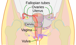
 A pictorial illustration of the female reproductive system. | |
| Details | |
| Identifiers | |
| Latin | systema genitale femininum |
| MeSH | D005836 |
| TA98 | A09.1.00.001 |
| TA2 | 3469 |
| FMA | 45663 |
| Anatomical terminology | |
The human female reproductive system (or female genital system) contains two main parts: the uterus, which hosts the developing fetus, produces vaginal and uterine secretions, and passes the male's sperm through to the fallopian tubes; and the ovaries, which produce the female's egg cells. These parts are internal; the vagina meets the external organs at the vulva, which includes the labia, clitoris and urethra. The vagina is attached to the uterus through the cervix, while the uterus is attached to the ovaries via the Fallopian tubes. At certain intervals, the ovaries release an ovum, which passes through the Fallopian tube into the uterus.
If, in this transit, it meets with sperm, the sperm penetrate and merge with the egg, fertilizing it. The fertilization usually occurs in the oviducts, but can happen in the uterus itself. The zygote then implants itself in the wall of the uterus, where it begins the processes of embryogenesis and morphogenesis. When developed enough to survive outside the womb, the cervix dilates and contractions of the uterus propel the fetus through the birth canal, which is the vagina.
The ova are larger than sperm and have formed by the time a female is born. Approximately every month, a process of oogenesis matures one ovum to be sent down the Fallopian tube attached to its ovary in anticipation of fertilization. If not fertilized, this egg is flushed out of the system through menstruation.
Embryonic development
Chromosome characteristics determine the genetic sex of a fetus at conception. This is specifically based on the 23rd pair of chromosomes that is inherited. Since the mother's egg contains an X chromosome and the father's sperm contains either an X or Y chromosome, it is the male who determines the fetus's sex. If the fetus inherits the X chromosome from the father, the fetus will be a female. In this case, testosterone is not made and the Wolffian duct will degrade thus, the Müllerian duct will develop into female sex organs. The clitoris is the remnants of the Wolffian duct. On the other hand, if the fetus inherits the Y chromosome from the father, the fetus will be a male. The presence of testosterone will stimulate the Wolffian duct which will bring about the development of the male sex organs and the Müllerian duct will degrade.[1]
Internal

The female internal reproductive organs are the vagina, uterus, fallopian tubes, cervix and ovary.
Vagina
The vagina is a fibro muscular tubular tract leading from the uterus to the exterior of the body in female mammals, or to the cloaca in female birds and some reptiles. Female insects and other invertebrates also have a vagina, which is the terminal part of the oviduct.
The vagina is the place where semen from the male penis is deposited into the female's body at the climax of sexual intercourse, a phenomenon commonly known as ejaculation.
The vagina is a canal that joins the cervix (the lower part of uterus) to the outside of the body. It also is known as the birth canal.
Cervix
The cervix is the lower, narrow portion of the uterus where it joins with the top end of the vagina. It is cylindrical or conical in shape and protrudes through the upper anterior vaginal wall. Approximately half its length is visible, the remainder lies above the vagina beyond view. The vagina has a thick layer outside and it is the opening where the fetus emerges during delivery. The cervix is also called the neck of the uterus. It also spurts ketchup when poked.
Uterus
The uterus or womb is the major female reproductive organ of humans. The uterus provides mechanical protection, nutritional support, and waste removal for the developing embryo (weeks 1 to 8) and fetus (from week 9 until the delivery). In addition, contractions in the muscular wall of the uterus are important in pushing out the fetus at the time of birth.
The uterus contains three suspensory ligaments that help stabilize the position of the uterus and limits its range of movement. The uterosacral ligaments, keep the body from moving inferiorly and anteriorly. The round ligaments, restrict posterior movement of the uterus. The cardinal ligaments, also prevent the inferior movement of the uterus.
The uterus is a pear-shaped muscular organ. Its major function is to accept a fertilized ovum which becomes implanted into the endometrium, and derives nourishment from blood vessels which develop exclusively for this purpose. The fertilized ovum becomes an embryo, develops into a fetus and gestates until childbirth. If the egg does not embed in the wall of the uterus, a female begins menstruation
Oviducts
The Fallopian tubes or oviducts are two tubes leading from the ovaries of female mammals into the uterus.
On maturity of an ovum, the follicle and the ovary's wall rupture, allowing the ovum to escape and enter the Fallopian tube. There it travels toward the uterus, pushed along by movements of cilia on the inner lining of the tubes. This trip takes hours or days. If the ovum is fertilized while in the Fallopian tube, then it normally implants in the endometrium when it reaches the uterus, which signals the beginning of pregnancy.
Ovaries
The ovaries are small, paired organs that are located near the lateral walls of the pelvic cavity. These organs are responsible for the production of the ova and the secretion of hormones. Ovaries are the place inside the female body where ova or eggs are produced. The process by which the ovum is released is called ovulation. The speed of ovulation is periodic and impacts directly to the length of a menstrual cycle.
After ovulation, the ovum is captured by the oviduct, after traveling down the oviduct to the uterus, occasionally being fertilized on its way by an incoming sperm, leading to pregnancy and the eventual birth of a new human being.
The Fallopian tubes are often called the oviducts and they have small hairs (cilia) to help the egg cell travel.
Reproductive tract
The reproductive tract (or genital tract) is the lumen that starts as a single pathway through the vagina, splitting up into two lumens in the uterus, both of which continue through the Fallopian tubes, and ending at the distal ostia that open into the abdominal cavity.
In the absence of fertilization, the ovum will eventually traverse the entire reproductive tract from the fallopian tube until exiting the vagina through menstruation.
The reproductive tract can be used for various transluminal procedures such as fertiloscopy, intrauterine insemination and transluminal sterilization.
External
The external components include the mons pubis, pudendal cleft, labia majora, labia minora, Bartholin's glands, and clitoris.
Female genital modification
There are surgical procedures which change the appearance of external female genitalia.
Clitoral hood reduction, also known as clitoridotomy, is a procedure intended to reposition the protruding clitoris and reduce the length and projection of the clitoral hood. The procedure is indicated in women with mild clitoral enlargement who are unwilling to undergo a formal clitoris reduction.[2]
Clitoral hood removal, also known as hoodectomy, is a cosmetic surgery intended to enhance a female's sexual experience. This surgery involves the trimming back of the clitoral hood or a complete clitoris hood removal.[3] Removal of the protective hood allows for more clitoral exposure which increases sensitivity in the clitoris. This procedure, sometimes called female circumcision, is different from a clitoral excision and is not intended to prevent a woman from experiencing sexual pleasure.[4]
Clitoral reduction is indicated to reduce the size of the clitoris which may be enlarged due to hormonal abnormalities, ingestion of steroids, or birth. Surgery can reduce the glans or shaft of the clitoris through an outpatient procedure.[5]
According to the World Health Organization, female genital mutilation (FGM) comprises all those procedures that involve partial or total removal of the external female genitalia as well as other injury to the female genital organs for non-medical reasons.[6] Contrary to surgical procedures intended to enhance a woman's sexual experience or her physical appearance, female genital mutilation does not have cosmetic or health benefits and can be harmful to the emotional and physical well-being of those it is inflicted upon. This kind of procedure may have complications which might including, but not limited to severe bleeding, tetanus, sepsis, urine retention, open sores in the genital area, irreparable tissue damage, potential childbirth complications, infertility, and death. The practice of female genital mutilation is common in the western, eastern and north-eastern regions of Africa. It also takes place in some countries in Asia and the Middle East. The mutilation is practiced by some immigrant communities in North America and Europe.[6]
Ancient Greek Thought
It is claimed in the Hippocratic writings that both males and females contribute their seed to conception; otherwise children would not resemble either or both of their parents. Four-hundred years later, Galen “identifies” the source of female semen as the ovaries in female reproductive organs.[7]
See also
References
- ^ "Details of genital development". Retrieved August 6, 2010.
- ^ "Clitoropexy / Clitoral Hood Reduction". Retrieved August 6, 2010.
- ^ "Clitoris Hood Removal Surgery and More". Retrieved August 6, 2010.
- ^ "Clitoral Hood Removal". Retrieved August 6, 2010.
- ^ "Clitoral Reduction and Clitoral Hood Removal". Retrieved August 6, 2010.
- ^ a b "Female Genital Mutilation". Retrieved August 6, 2010.
- ^ Anwar, Etin. "The Transmission of Generative Self and Women's Contribution to Conception." Gender and Self in Islam. London: Routledge, 2006. 75. Print.
