
Medial rectus muscle
| Medial rectus | |
|---|---|
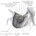
 Rectus muscles: 2 = superior, 3 = inferior, 4 = medial, 5 = lateral Oblique muscles: 6 = superior, 8 = inferior Other muscle: 9 = levator palpebrae superioris Other structures: 1 = Annulus of Zinn, 7 = Trochlea, 10 = Superior tarsus, 11 = Sclera, 12 = Optic nerve | |
 Figure showing the mode of innervation of the Recti medialis and lateralis of the eye. | |
| Details | |
| Origin | annulus of Zinn at the orbital apex |
| Insertion | 5.5 mm medial to the limbus |
| Nerve | inferior division of the oculomotor nerve |
| Actions | adducts the eyeball (makes it move inwards) |
| Identifiers | |
| Latin | musculus rectus medialis bulbi |
| TA98 | A15.2.07.012 |
| TA2 | 2044 |
| FMA | 49037 |
| Anatomical terms of muscle | |
The medial rectus muscle is a muscle in the orbit.
As with most of the muscles of the orbit, it is innervated by the inferior division of the oculomotor nerve (Cranial Nerve III).
This muscle shares an origin with several other extrinsic eye muscles, the anulus tendineus, or common tendon.
It is the largest of the extraocular muscles and its only action is adduction of the eyeball. Its function is to bring the pupil closer to the midline of the body. It is tested clinically by asking the patient to look medially.
Additional images
-
 Eye movement of medial rectus muscle, superior view.
 -
 Horizontal section of the eyeball.
 -
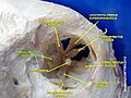
 Dissection showing origins of right ocular muscles, and nerves entering by the superior orbital fissure.
 -

-
 Medial rectus muscle
 -
 Medial rectus muscle
 -
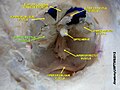
 Extrinsic eye muscle. Nerves of orbita. Deep dissection.
 -
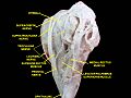 Extrinsic eye muscle. Nerves of orbita. Deep dissection.
 -
 Extrinsic eye muscle. Nerves of orbita. Deep dissection.
 -
 Extrinsic eye muscle. Nerves of orbita. Deep dissection.
 -
 Extrinsic eye muscle. Nerves of orbita. Deep dissection.
 -
 Extrinsic eye muscle. Nerves of orbita. Deep dissection.


External links
- Anatomy figure: 29:01-06 at Human Anatomy Online, SUNY Downstate Medical Center
- lesson3 at The Anatomy Lesson by Wesley Norman (Georgetown University) (orbit4)
- Diagram at howstuffworks.com
This muscle article is a stub. You can help Wikipedia by expanding it. |
This article about the eye is a stub. You can help Wikipedia by expanding it. |