
Superior rectus muscle
| Superior rectus | |
|---|---|
 View of the eye from above, showing the action of the superior rectus muscle. | |
| Details | |
| Origin | annulus of Zinn at the orbital apex |
| Insertion | 7.5 mm superior to the limbus |
| Nerve | oculomotor nerve |
| Actions | elevates, intorsion, and rotates medially the eye |
| Identifiers | |
| Latin | musculus rectus superior bulbi |
| TA98 | A15.2.07.010 |
| TA2 | 2042 |
| FMA | 49035 |
| Anatomical terms of muscle | |
The superior rectus muscle is a muscle in the orbit. It is one of the extraocular muscles. It is innervated by the superior division of the oculomotor nerve (Cranial Nerve III). In the primary position (looking straight ahead), the superior rectus muscle's primary function is elevation, although it also contributes to intorsion and adduction.

Structure
Function
It elevates, adducts, and helps intort (rotate medially) the eye.
Clinical significance
Testing
The superior rectus muscle is the only muscle that is capable of elevating the eye when it is in a fully abducted position.[1]
Additional images
This gallery of anatomic features needs cleanup to abide by the medical manual of style. |
-
 The right eye in sagittal section, showing the fascia bulbi (semidiagrammatic).
 -
 Superior rectus muscle
 -
 Superior rectus muscle
 -
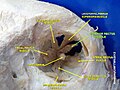
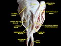
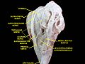 Extrinsic eye muscle. Nerves of orbita. Deep dissection.
 -
 Extrinsic eye muscle. Nerves of orbita. Deep dissection.
 -
 Extrinsic eye muscle. Nerves of orbita. Deep dissection.
 -
 Extrinsic eye muscle. Nerves of orbita. Deep dissection.
 -
 Extrinsic eye muscle. Nerves of orbita. Deep dissection.
 -
 Extrinsic eye muscle. Nerves of orbita. Deep dissection.
 -
 Extrinsic eye muscle. Nerves of orbita. Deep dissection.
 -
 Extrinsic eye muscle. Nerves of orbita. Deep dissection.


References
- ^ "Eye Theory". Cim.ucdavis.edu. Retrieved 2012-12-02.
External links
- Anatomy figure: 29:01-02 at Human Anatomy Online, SUNY Downstate Medical Center
- "Diagram". Archived from the original on March 25, 2010.