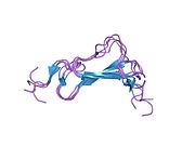
TGF alpha
Template:PBB Transforming growth factor alpha (TGF-α) is a protein that in humans is encoded by the TGFA gene.[1] As a member of the epidermal growth factor (EGF) family, TGF-α is a mitogenic polypeptide.[2] The protein becomes activated when binding to receptors capable of protein kinase activity for cellular signaling.
TGF-α is a transforming growth factor that is a ligand for the epidermal growth factor receptor, which activates a signaling pathway for cell proliferation, differentiation and development. This protein may act as either a transmembrane-bound ligand or a soluble ligand. This gene has been associated with many types of cancers, and it may also be involved in some cases of cleft lip/palate.[1]
Synthesis
TGF-α is synthesized internally as part of a 160 (human) or 159 (rat) amino acid transmembrane precursor.[3] The precursor is composed of an extracellular domain containing a hydrophobic transmembrane domain, 50 amino acids of TGF-α, and a 35-residue-long cytoplasmic domain.[3] In its smallest form TGF-α has six cysteines linked together via three disulfide bridges. Collectively all members of the EGF/TGF-α family share this structure. The protein, however, is not directly related to TGF-β. In the stomach, TGF-α is manufactured within the normal gastric mucosa.[4] TGF-α has been shown to inhibit gastric acid secretion.[4]
Limited success has resulted from attempts to synthesize of a reductant molecule to TGF-α that displays a similar biological profile.[5]
Synthesis in the stomach
In the stomach, TGF-α is manufactured within the normal gastric mucosa.[4] TGF-α has been shown to inhibit gastric acid secretion.[4]
Function
TGF-α can be produced in macrophages, brain cells, and keratinocytes. TGF-α induces epithelial development. Considering that TGF-α is a member of the EGF family, the biological actions of TGF-α and EGF are similar. For instance, TGF-α and EGF bind to the same receptor. When TGF-α binds to EGFR it can initiate multiple cell proliferation events.[5] Cell proliferation events that involve TGF-α bound to EGFR include wound healing and embryogenesis. TGF-α is also involved in tumerogenesis and believed to promote angiogenesis.[3]
TGFα has also been shown to stimulate neural cell proliferation in the adult injured brain.[6]
TGF-α receptor
A 170-kDa glycosylated protein known as the EGF receptor binds to TGF-α allowing the polypeptide to function in various signaling pathways.[2] The EGF receptor is characterized by having an extracellular domain that has numerous amino acid motifs. EGFR is essential for a single transmembrane domain, an intracellular domain (containing tyrosine kinase activity), and ligand recognition.[2] As a membrane anchored-growth factor, TGF-α can be cleaved from an integral membrane glycoprotein via a protease.[3] Soluble forms of TGF-α resulting from the cleavage have the capacity to activate EGFR. EGFR can be activated from a membrane-anchored growth factor as well.
When TGF-α binds to EGFR it dimerizes triggering phosphorylation of a protein-tyrosine kinase. The activity of protein-tyrosine kinase causes an autophosphorylation to occur among several tyrosine residues within EGFR, influencing activation and signaling of other proteins that interact in many signal transduction pathways.

Animal studies
In an animal model of Parkinson's disease where dopaminergic neurons have been damaged by 6-hydroxydopamine, infusion of TGF-α into the brain caused an increase in the number of neuronal precursor cells.[6] However TGF-α treatment did not result in neurogenesis dopaminergic neurons.[7]
Human studies
TGF-α and the neuroendocrine system
The EGF/TGF-α family has been shown to regulate luteinizing hormone-releasing hormone (LHRH) through a glial-neuronal interactive process.[2] Produced in hypothalamic astrocytes, TGF-α indirectly stimulates LHRH release through various intermediates. As a result, TGF-α is a physiological component essential to the initiation process of female puberty.[2]
TGF-α and the suprachiasmatic nucleus
TGF-α has also been observed to be highly expressed in the suprachiasmatic nucleus (SCN) (5). This finding suggests a role for EGFR signaling in the regulation of CLOCK and circadian rhythms within the SCN.[8] Similar studies have shown that when injected into the third ventricle TGF-α can suppress circadian locomotor behavior along with drinking or eating activities.[8]
TGF-α and tumors
Its potential use as a prognostic biomarker in various tumors, like gastric carcinoma .[9] or melanoma has been suggested.[10]
Interactions
TGF alpha has been shown to interact with GORASP1[11] and GORASP2.[11]
See also
References
- ^ a b "Entrez Gene: TGFA transforming growth factor alpha".
- ^ a b c d e Ojeda, S.; Ma, Y.; Rage, F. The transforming growth factor alpha gene family is involved in the neuroendocrine control of mammalian puberty. Mol. Psychiatry 1997, 2, 355.
- ^ a b c d Ferrer, I.; Alcantara, S.; Ballabriga, J.; Olive, M.; Blanco, R.; Rivera, R.; Carmona, M.; Berruezo, M.; Pitarch, S.; Planas, A. Transforming growth factor- α (TGF-α) and epidermal growth factor-receptor (EGF-R) immunoreactivity in normal and pathologic brain. Prog. Neurobiol. 1996, 49, 99.
- ^ a b c d Coffey, R.; Gangarosa, L.; Damstrup, L.; Dempsey, P. Basic actions of transforming growth factor- α and related peptides. Eur. J. Gastroen. Hepat. 1995, 7, 923.
- ^ a b McInnes, C; Wang, J; Al Moustafa, AE; Yansouni, C; O'Connor-McCourt, M; Sykes, BD (1998). "Structure-based minimization of transforming growth factor-alpha (TGF-alpha) through NMR analysis of the receptor-bound ligand. Design, solution structure, and activity of TGF-alpha 8-50"". J. Biol. Chem. 273 (42): 27357–63. doi:10.1074/jbc.273.42.27357.
{{cite journal}}: CS1 maint: unflagged free DOI (link) - ^ a b Fallon J, Reid S, Kinyamu R, Opole I, Opole R, Baratta J, Korc M, Endo TL, Duong A, Nguyen G, Karkehabadhi M, Twardzik D, Patel S, Loughlin S (2000). "In vivo induction of massive proliferation, directed migration, and differentiation of neural cells in the adult mammalian brain". Proceedings of the National Academy of Sciences of the United States of America. 97 (26): 14686–91. doi:10.1073/pnas.97.26.14686. PMC 18979. PMID 11121069.
{{cite journal}}: CS1 maint: multiple names: authors list (link) - ^ Cooper O, Isacson O (October 2004). "Intrastriatal transforming growth factor alpha delivery to a model of Parkinson's disease induces proliferation and migration of endogenous adult neural progenitor cells without differentiation into dopaminergic neurons". J. Neurosci. 24 (41): 8924–31. doi:10.1523/JNEUROSCI.2344-04.2004. PMC 2613225. PMID 15483111.
- ^ a b Hao, H.; Schwaber, J. Epidermal growth factor receptor induced Erk phosphorylation in the suprachiasmatic nucleus. Brain Res. 2006, 1088, 45.
- ^ Fanelli MF (Aug 2012). "The influence of transforming growth factor-α, cyclooxygenase-2, matrix metalloproteinase (MMP)-7, MMP-9 and CXCR4 proteins involved in epithelial-mesenchymal transition on overall survival of patients with gastric cancer". Histopathology. 61 (2): 153–61. doi:10.1111/j.1365-2559.2011.04139.x. PMID 22582975.
- ^ Tarhini AA (Jan 2014). "A four-marker signature of TNF-RII, TGF-α, TIMP-1 and CRP is prognostic of worse survival in high-risk surgically resected melanoma." J Transl Med. 12. doi:10.1186/1479-5876-12-19. PMC 3909384. PMID 24457057.
{{cite journal}}: CS1 maint: unflagged free DOI (link) - ^ a b Barr FA, Preisinger C, Kopajtich R, Körner R (December 2001). "Golgi matrix proteins interact with p24 cargo receptors and aid their efficient retention in the Golgi apparatus". J. Cell Biol. 155 (6): 885–91. doi:10.1083/jcb.200108102. PMC 2150891. PMID 11739402.
{{cite journal}}: CS1 maint: multiple names: authors list (link)
Further reading
- Luetteke NC, Lee DC (1991). "Transforming growth factor alpha: expression, regulation and biological action of its integral membrane precursor". Semin. Cancer Biol. 1 (4): 265–75. PMID 2103501.
- Greten FR, Wagner M, Weber CK, Zechner U, Adler G, Schmid RM (2002). "TGF alpha transgenic mice. A model of pancreatic cancer development". Pancreatology. 1 (4): 363–8. doi:10.1159/000055835. PMID 12120215.
{{cite journal}}: CS1 maint: multiple names: authors list (link) - Vieira AR (2006). "Association between the transforming growth factor alpha gene and nonsyndromic oral clefts: a HuGE review". Am. J. Epidemiol. 163 (9): 790–810. doi:10.1093/aje/kwj103. PMID 16495466.
- Nasim MM, Thomas DM, Alison MR, Filipe MI (1992). "Transforming growth factor alpha expression in normal gastric mucosa, intestinal metaplasia, dysplasia and gastric carcinoma--an immunohistochemical study". Histopathology. 20 (4): 339–43. doi:10.1111/j.1365-2559.1992.tb00991.x. PMID 1577411.
{{cite journal}}: CS1 maint: multiple names: authors list (link) - Thomas DM, Nasim MM, Gullick WJ, Alison MR (1992). "Immunoreactivity of transforming growth factor alpha in the normal adult gastrointestinal tract". Gut. 33 (5): 628–31. doi:10.1136/gut.33.5.628. PMC 1379291. PMID 1612477.
{{cite journal}}: CS1 maint: multiple names: authors list (link) - Bean MF, Carr SA (1992). "Characterization of disulfide bond position in proteins and sequence analysis of cystine-bridged peptides by tandem mass spectrometry". Anal. Biochem. 201 (2): 216–26. doi:10.1016/0003-2697(92)90331-Z. PMID 1632509.
- Lei ZM, Rao CV (1992). "Expression of epidermal growth factor (EGF) receptor and its ligands, EGF and transforming growth factor-alpha, in human fallopian tubes". Endocrinology. 131 (2): 947–57. doi:10.1210/en.131.2.947. PMID 1639032.
- Werner S, Roth WK, Bates B, Goldfarb M, Hofschneider PH (1991). "Fibroblast growth factor 5 proto-oncogene is expressed in normal human fibroblasts and induced by serum growth factors". Oncogene. 6 (11): 2137–44. PMID 1658709.
{{cite journal}}: CS1 maint: multiple names: authors list (link) - Saeki T, Cristiano A, Lynch MJ, Brattain M, Kim N, Normanno N, Kenney N, Ciardiello F, Salomon DS (1992). "Regulation by estrogen through the 5'-flanking region of the transforming growth factor alpha gene". Mol. Endocrinol. 5 (12): 1955–63. doi:10.1210/mend-5-12-1955. PMID 1791840.
{{cite journal}}: CS1 maint: multiple names: authors list (link) - Harvey TS, Wilkinson AJ, Tappin MJ, Cooke RM, Campbell ID (1991). "The solution structure of human transforming growth factor alpha". Eur. J. Biochem. 198 (3): 555–62. doi:10.1111/j.1432-1033.1991.tb16050.x. PMID 2050136.
{{cite journal}}: CS1 maint: multiple names: authors list (link) - Kline TP, Brown FK, Brown SC, Jeffs PW, Kopple KD, Mueller L (1991). "Solution structures of human transforming growth factor alpha derived from 1H NMR data". Biochemistry. 29 (34): 7805–13. doi:10.1021/bi00486a005. PMID 2261437.
{{cite journal}}: CS1 maint: multiple names: authors list (link) - Jakowlew SB, Kondaiah P, Dillard PJ, Sporn MB, Roberts AB (1989). "A novel low molecular weight ribonucleic acid (RNA) related to transforming growth factor alpha messenger RNA". Mol. Endocrinol. 2 (11): 1056–63. doi:10.1210/mend-2-11-1056. PMID 2464748.
{{cite journal}}: CS1 maint: multiple names: authors list (link) - Jakobovits EB, Schlokat U, Vannice JL, Derynck R, Levinson AD (1989). "The human transforming growth factor alpha promoter directs transcription initiation from a single site in the absence of a TATA sequence". Mol. Cell. Biol. 8 (12): 5549–54. PMC 365660. PMID 2907605.
{{cite journal}}: CS1 maint: multiple names: authors list (link) - Tricoli JV, Nakai H, Byers MG, Rall LB, Bell GI, Shows TB (1986). "The gene for human transforming growth factor alpha is on the short arm of chromosome 2". Cytogenet. Cell Genet. 42 (1–2): 94–8. doi:10.1159/000132258. PMID 3459638.
{{cite journal}}: CS1 maint: multiple names: authors list (link) - Lee DC, Rose TM, Webb NR, Todaro GJ (1985). "Cloning and sequence analysis of a cDNA for rat transforming growth factor-alpha". Nature. 313 (6002): 489–91. doi:10.1038/313489a0. PMID 3855503.
{{cite journal}}: CS1 maint: multiple names: authors list (link) - Derynck R, Roberts AB, Winkler ME, Chen EY, Goeddel DV (1984). "Human transforming growth factor-alpha: precursor structure and expression in E. coli". Cell. 38 (1): 287–97. doi:10.1016/0092-8674(84)90550-6. PMID 6088071.
{{cite journal}}: CS1 maint: multiple names: authors list (link) - Ogbureke KU, MacDaniel RK, Jacob RS, Durban EM (1995). "Distribution of immunoreactive transforming growth factor-alpha in non-neoplastic human salivary glands". Histol. Histopathol. 10 (3): 691–6. PMID 7579819.
{{cite journal}}: CS1 maint: multiple names: authors list (link) - Walz TM, Malm C, Nishikawa BK, Wasteson A (1995). "Transforming growth factor-alpha (TGF-alpha) in human bone marrow: demonstration of TGF-alpha in erythroblasts and eosinophilic precursor cells and of epidermal growth factor receptors in blastlike cells of myelomonocytic origin". Blood. 85 (9): 2385–92. PMID 7727772.
{{cite journal}}: CS1 maint: multiple names: authors list (link) - Patel B, Hiscott P, Charteris D, Mather J, McLeod D, Boulton M (1994). "Retinal and preretinal localisation of epidermal growth factor, transforming growth factor alpha, and their receptor in proliferative diabetic retinopathy". The British journal of ophthalmology. 78 (9): 714–8. doi:10.1136/bjo.78.9.714. PMC 504912. PMID 7947554.
{{cite journal}}: CS1 maint: multiple names: authors list (link)
External links
- Transforming+Growth+Factor+alpha at the U.S. National Library of Medicine Medical Subject Headings (MeSH)
This article incorporates text from the United States National Library of Medicine, which is in the public domain.


