
Heterochromia iridum
| Heterochromia iridum | |
|---|---|
| Specialty | Ophthalmology |
In anatomy, heterochromia (Greek: heteros 'different' + chroma 'color'[1]) refers to a difference in coloration, usually of the iris but also of hair or skin. Heterochromia is a result of the relative excess or lack of melanin (a pigment). It may be inherited, or caused by genetic mosaicism, chimerism, disease, or injury.[2]
Heterochromia of the eye (heterochromia iridis or heterochromia iridum; the common incorrect form "heterochromia iridium" is not correct Latin) is of two kinds. In complete heterochromia, one iris is a different color from the other. In partial heterochromia or sectoral heterochromia, part of one iris is a different color from its remainder.
Eye color, specifically the color of the irises, is determined primarily by the concentration and distribution of melanin.[3][4][5] The affected eye may be hyperpigmented (hyperchromic) or hypopigmented (hypochromic).[6] In humans, usually, an excess of melanin indicates hyperplasia of the iris tissues, whereas a lack of melanin indicates hypoplasia.
Classification
Heterochromia is classified primarily by onset: as either genetic or acquired. Although a distinction is frequently made between heterochromia that affects an eye completely or only partially (sectoral heterochromia), it is often classified as either genetic (due to mosaicism or congenital) or acquired, with mention as to whether the affected iris or portion of the iris is darker or lighter. [7] Most cases of heterochromia are hereditary, caused by a disease or syndrome, or due to an injury. Sometimes one eye may change color following certain diseases or injuries.[8]

Congenital heterochromia
Heterochromia that is congenital is usually inherited as an autosomal dominant trait.
Abnormal iris darker
- Lisch nodules – iris hamartomas seen in neurofibromatosis.
- Ocular melanosis – a condition characterized by increased pigmentation of the uveal tract, episclera, and anterior chamber angle.
- Oculodermal melanocytosis (nevus of Ota)[6]
- Pigment dispersion syndrome – a condition characterized by loss of pigmentation from the posterior iris surface which is disseminated intraocularly and deposited on various intraocular structures, including the anterior surface of the iris.
- Sturge-Weber syndrome – a syndrome characterized by a port-wine stain nevus in the distribution of the trigeminal nerve, ipsilateral leptomeningeal angiomas (leptomaningiomas) with intracranial calcification and neurologic signs, and angioma of the choroid, often with secondary glaucoma.[9][10]
Abnormal iris lighter
- Simple heterochromia – a rare condition characterized by the absence of other ocular or systemic problems. The lighter eye is typically regarded as the affected eye as it usually shows iris hypoplasia. It may affect an iris completely or only partially.
- Congenital Horner's syndrome[11] – sometimes inherited, although usually acquired
- Waardenburg's syndrome[11] – a syndrome in which heterochromia is expressed as a bilateral iris hypochromia in some cases. A Japanese review of 11 albino children with the disorder found that all had sectoral/partial heterochromia.[12]
- Piebaldism – similar to Waardenburg's syndrome, a rare disorder of melanocyte development characterized by a white forelock and multiple symmetrical hypopigmented or depigmented macules.
- Hirschsprung's disease – a bowel disorder associated with heterochromia in the form of a sector hypochromia. The affected sectors have been shown to have reduced numbers of melanocytes and decreased stromal pigmentation.[13]
- Incontinentia pigmenti[6]
- Parry-Romberg syndrome[6]
Acquired heterochromia
Heterochromia that is acquired is usually due to injury, inflammation, the use of certain eyedrops,[14] or tumors.
Abnormal iris darker
- Deposition of material
- Siderosis – iron deposition within ocular tissues due to a penetrating injury and a retained iron-containing, intraocular foreign body.
- Hemosiderosis – long standing hyphema (blood in the anterior chamber) following blunt trauma to the eye may lead to iron deposition from blood products
- Certain eyedrops – prostaglandin analogues (latanoprost, isopropyl unoprostone, travoprost, and bimatoprost) are used topically to lower intraocular pressure in glaucoma patients. A concentric heterochromia has developed in some patients applying these drugs. The stroma around the iris sphincter muscle becomes darker than the peripheral stroma. A stimulation of melanin synthesis within iris melanocytes has been postulated.
- Neoplasm – Nevi and melanomatous tumors.
- Iridocorneal endothelium syndrome[6]
- Iris ectropion syndrome[6]
Abnormal iris lighter
- Fuchs heterochromic iridocyclitis – a condition characterized by a low grade, asymptomatic uveitis in which the iris in the affected eye becomes hypochromic and has a washed-out, somewhat moth eaten appearance. The heterochromia can be very subtle, especially in patients with lighter colored irides. It is often most easily seen in daylight. The prevalence of heterochromia associated with Fuchs has been estimated in various studies[15][16][17] with results suggesting that there is more difficulty recognizing iris color changes in dark-eyed individuals.[17][18]
- Acquired Horner's syndrome – usually acquired, as in neuroblastoma,[19] although sometimes inherited.
- Neoplasm – Melanomas can also be very lightly pigmented, and a lighter colored iris may be a rare manifestation of metastatic disease to the eye.
Heterochromia has also been observed in those with Duane syndrome.[20][21]
Central heterochromia

Central heterochromia is an eye condition where there are two colors in the same iris; the central (pupillary) zone of the iris is a different color than the mid-peripheral (ciliary) zone, with the true iris color being the outer color. [22]
Eye color is determined primarily by the concentration and distribution of melanin within the iris tissues. Although the processes determining eye color are not fully understood, it is known that inherited eye color is determined by multiple genes. Environmental or acquired factors can alter these inherited traits.[3]
The human iris can be seen in a number of various colors. There are three true colors in human eyes that determine the outward appearance: brown, yellow, and grey. The amount of each color an individual has determines the appearance of the eye color.[23]
Eyes displaying central heterochromia are often referred to as "cat eyes" because of their multi-colored iris. Central heterochromia appears to be prevalent in irises containing low amounts of melanin.[24]
A famous case of a person with central heterochromia was Baroness Rozsika Edle von Wertheimstein, whose daughter wrote: "She was a very beautiful woman... She had dark, dark brown eyes, but each eye had a purple ring to it, about a quarter of an inch of purple around these dark brown eyes."[25]
Notable people with heterochromia
In other organisms
Although infrequently seen in humans, complete heterochromia is more frequently observed in other species, where it almost always involves one blue eye. The blue eye occurs within a white spot, where melanin is absent from the skin and hair (see Leucism). These species include the cat, particularly breeds such as Turkish Van, Turkish Angora, Khao Manee and (rarely) Japanese Bobtail. These so-called odd-eyed cats are white, or mostly white, with one normal eye (copper, orange, yellow, green), and one blue eye. Among dogs, complete heterochromia is seen often in the Siberian Husky and few other breeds, usually Australian Shepherd and Catahoula Leopard Dog. Horses with complete heterochromia have one brown and one white, gray, or blue eye - complete heterochromia is more common in horses with pinto coloring. Complete heterochromia occurs also in cattle and even water buffalo.[26] It can also be seen in ferrets with Waardenburg Syndrome, although it can be very hard to tell at times as the eye color is often a midnight blue.
Sectoral heterochromia, usually sectoral hypochromia, is often seen in dogs, specifically in breeds with merle coats. These breeds include the Australian Shepherd, Border Collie, Collie, Shetland Sheepdog, Welsh Corgi, Pyrenean Shepherd, Mudi, Beauceron, Catahoula Cur, Dunker, Great Dane, Dachshund and Chihuahua. It also occurs in certain breeds that do not carry the merle trait, such as the Siberian Husky.
Gallery
-
Complete heterochromia in a teenager, which also has anisocoria.
-
 A young adult exhibiting sectoral heterochromia in the form of an orange segment in her right, blue eye. The individual's mother exhibited a similar orange segment in her left eye, although her iris color was green.
 -
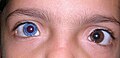 Heterochromia in a child.
 -
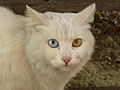 Complete heterochromia in a cat: one eye blue, one yellow. The yellow eye has what looks like central heterochromia, as the outside of the eye is yellow, and the iris is green.
 -
 Complete heterochromia in a Siberian Husky: one eye blue, one eye brown.
 -
 Sectoral hypochromia in a blue merle Border Collie.
 -
 A cat with complete heterochromia.
 -
 Example of central heterochromia showing an orange to blue iris.
 -
An example of a green eye with a brown section.
-
A spotted kitten with sectoral heterochromia


See also
- Brushfield spots
- Coloboma
- Erythrism
- Leucism
- List of systemic diseases with ocular manifestations
- Piebaldism
References
- ^ http://medical-dictionary.thefreedictionary.com/heterochromia+iridis
- ^ Imesch PD, Wallow IH, Albert DM (1997). "The color of the human eye: a review of morphologic correlates and of some conditions that affect iridial pigmentation throughout life". Surv Ophthalmol. 41 (Suppl 2): S117–23. doi:10.1016/S0039-6257(97)80018-5. PMID 9154287.
{{cite journal}}: Unknown parameter|month=ignored (help)CS1 maint: multiple names: authors list (link) - ^ a b Wielgus AR, Sarna T (2005). "Melanin in human irides of different color and age of donors". Pigment Cell Res. 18 (6): 454–64. doi:10.1111/j.1600-0749.2005.00268.x. PMID 16280011.
{{cite journal}}: Unknown parameter|month=ignored (help) - ^ Prota G, Hu DN, Vincensi MR, McCormick SA, Napolitano A (1998). "Characterization of melanins in human irides and cultured uveal melanocytes from eyes of different colors". Exp Eye Res. 67 (3): 293–9. doi:10.1006/exer.1998.0518. PMID 9778410.
{{cite journal}}: Unknown parameter|month=ignored (help)CS1 maint: multiple names: authors list (link) - ^ "All About Eye Color" from Larry Bickford
- ^ a b c d e f g h i Loewenstein, John; Scott Lee (2004). Ophthalmology: Just the Facts. New York: McGraw-Hill. ISBN 0-07-140332-9.
{{cite book}}: CS1 maint: multiple names: authors list (link) - ^ Swann P. "Heterochromia." Optometry Today. January 29, 1999. Retrieved November 1, 2006.
- ^ http://www.nlm.nih.gov/medlineplus/ency/article/003319.htm
- ^ van Emelen C, Goethals M, Dralands L, Casteels I (2000). "Treatment of glaucoma in children with Sturge-Weber syndrome". J Pediatr Ophthalmol Strabismus. 37 (1): 29–34. PMID 10714693.
{{cite journal}}: Unknown parameter|month=ignored (help)CS1 maint: multiple names: authors list (link) - ^ "Sturge-Weber syndrome: Definition and Much More from Answers.com". Answers.com<!. Retrieved 2009-11-19.
- ^ a b Wallis DH, Granet DB, Levi L (2003). "When the darker eye has the smaller pupil". J Aapos. 7 (3): 215–6. doi:10.1016/S1091-8531(02)42020-4. PMID 12825064.
{{cite journal}}: Unknown parameter|month=ignored (help)CS1 maint: multiple names: authors list (link) - ^ Ohno N, Kiyosawa M, Mori H, Wang WF, Takase H, Mochizuki M (2003). "Clinical findings in Japanese patients with Waardenburg syndrome type 2". Jpn J Ophthalmol. 47 (1): 77–84. doi:10.1016/S0021-5155(02)00629-9. PMID 12586183.
{{cite journal}}: Unknown parameter|month=ignored (help)CS1 maint: multiple names: authors list (link) - ^ Brazel SM, Sullivan TJ, Thorner PS, Clarke MP, Hunter WS, Morin JD (1992). "Iris sector heterochromia as a marker for neural crest disease". Arch Ophthalmol. 110 (2): 233–5. PMID 1736874.
{{cite journal}}: Unknown parameter|month=ignored (help)CS1 maint: multiple names: authors list (link) - ^ Liu CSC (1999). "A case of acquired iris depigmentation as a possible complication of levobunolol eye drops". British Journal of Ophthalmology. 83 (12). doi:10.1136/bjo.83.12.1403c.
{{cite journal}}: Unknown parameter|month=ignored (help) - ^ Yang P, Fang W, Jin H, Li B, Chen X, Kijlstra A (2006). "Clinical features of Chinese patients with Fuchs' syndrome". Ophthalmology. 113 (3): 473–80. doi:10.1016/j.ophtha.2005.10.028. PMID 16458965.
{{cite journal}}: Unknown parameter|month=ignored (help)CS1 maint: multiple names: authors list (link) - ^ Arellanes-Garcia L, del Carmen Preciado-Delgadillo M, Recillas-Gispert C (2002). "Fuchs' heterochromic iridocyclitis: clinical manifestations in dark-eyed Mexican patients". Ocul Immunol Inflamm. 10 (2): 125–31. doi:10.1076/ocii.10.2.125.13976. PMID 12778348.
{{cite journal}}: Unknown parameter|month=ignored (help)CS1 maint: multiple names: authors list (link) - ^ a b Tabbut BR, Tessler HH, Williams D (1988). "Fuchs' heterochromic iridocyclitis in blacks". Arch Ophthalmol. 106 (12): 1688–90. PMID 3196209.
{{cite journal}}: Unknown parameter|month=ignored (help)CS1 maint: multiple names: authors list (link) - ^ Bloch-Michel E (1983). "Fuchs heterochromic cyclitis: current concepts". J Fr Ophtalmol. (in French). 6 (10): 853–8. PMID 6368659.
- ^ Mehta K, Haller JO, Legasto AC (2003). "Imaging neuroblastoma in children". Crit Rev Comput Tomogr. 44 (1): 47–61. doi:10.1080/10408370390808469. PMID 12627783.
{{cite journal}}: CS1 maint: multiple names: authors list (link) - ^ Khan AO, Aldamesh M (2006). "Bilateral Duane syndrome and bilateral aniridia". J Aapos. 10 (3): 273–4. doi:10.1016/j.jaapos.2006.02.002. PMID 16814183.
{{cite journal}}: Unknown parameter|month=ignored (help) - ^ Shauly Y, Weissman A, Meyer E (1993). "Ocular and systemic characteristics of Duane syndrome". J Pediatr Ophthalmol Strabismus. 30 (3): 178–83. PMID 8350229.
{{cite journal}}: Unknown parameter|month=ignored (help)CS1 maint: multiple names: authors list (link) - ^ HeterochromiaCentral.com - What Is Central Heterochromia?
- ^ Seddon JM, Sahagian CR, Glynn RJ, Sperduto RD, Gragoudas ES (1990). "Evaluation of an iris color classification system". Invest Ophthalmol Vis Sci. 31 (8): 1592–8. PMID 2201662.
{{cite journal}}: Unknown parameter|month=ignored (help)CS1 maint: multiple names: authors list (link) - ^ "Key Ocular Signs for Screening". Milesresearch.com. Retrieved 2009-11-19.
- ^ Dame Miriam Rothschild, by Naomi Gryn, Jewish Quarterly, Spring 2004, page 54
- ^ Misk NA, Semieka MA, Fathy A (1998). "Heterochromia iridis in water buffaloes (Bubalus bubalis)". Vet Ophthalmol. 1 (4): 195–201. doi:10.1046/j.1463-5224.1998.00036.x. PMID 11397231.
{{cite journal}}: CS1 maint: multiple names: authors list (link)