Skin condition: Difference between revisions
m →Distribution: added tags |
|||
| Line 34: | Line 34: | ||
* '''Telangiectasia''' - A telangiectasia represents an enlargement of superficial blood vessels to the point of being visible.<ref name="isbn0-7216-8256-1" /> |
* '''Telangiectasia''' - A telangiectasia represents an enlargement of superficial blood vessels to the point of being visible.<ref name="isbn0-7216-8256-1" /> |
||
* '''Burrow''' - A burrow appears as a slightly elevated, grayish, tortuous line in the skin, and is caused by burrowing organisms.<ref name="isbn0-7216-8256-1" /><ref name="Andrews" /> |
* '''Burrow''' - A burrow appears as a slightly elevated, grayish, tortuous line in the skin, and is caused by burrowing organisms.<ref name="isbn0-7216-8256-1" /><ref name="Andrews" /> |
||
[http://major-diseases2011.blogspot.com/ Major Skin Diseases] |
|||
==== Secondary lesions ==== |
==== Secondary lesions ==== |
||
Revision as of 15:32, 15 June 2011
There are many conditions of or affecting the human integumentary system—the organ system that comprises the entire surface of the body and includes skin, hair, nails, and related muscle and glands.[1]
History
In 1572, Geronimo Mercuriali of Forlì, Italy, completed De morbis cutaneis (translated "On the diseases of the skin"). It is considered the first scientific work dedicated to dermatology.
Epidemiology
In World War I, over two million days of service are estimated to have been lost by reason of skin diseases alone.[2]
Approach to diagnoses
The physical examination of the skin and its appendages, as well as the mucous membranes, forms the cornerstone of an accurate diagnosis of cutaneous conditions.[3] Most of these conditions present with cutaneous surface changes term "lesions," which have more or less distinct characteristics.[4] Often proper examination will lead the physician to obtain appropriate historical information and/or laboratory tests that are able to confirm the diagnosis.[3] Upon examination, the important clinical observations are the (1) morphology, (2) configuration, and (3) distribution of the lesion(s).[3] With regard to morphology, the initial lesion that characterizes a condition is known as the "primary lesion," and identification of such a lesions is the most important aspect of the cutaneous examination.[4] Over time, these primary lesions may continue to develop or be modified by regression or trauma, producing "secondary lesions."[1] However, with that being stated, the lack of standardization of basic dermatologic terminology has been one of the principal barriers to successful communication among physicians in describing cutaneous findings.[5] Nevertheless, there are some commonly accepted terms used to describe the macroscopic morphology, configuration, and distribution of skin lesions, which are listed below.[4]
Morphology
Primary lesions


- Macule - A macule is a change in surface color, without elevation or depression and, therefore, nonpalpable, well or ill-defined,[6] variously sized, but generally considered less than either 5[6] or 10mm in diameter at the widest point.[4]
- Patch - A patch is a large macule equal to or greater than either 5 or 10mm,[4] depending on one's definition of a macule.[1] Patches may have some subtle surface change, such as a fine scale or wrinkling, but although the consistency of the surface is changed, the lesion itself is not palpable.[3]
- Papule - A papule is a circumscribed, solid elevation of skin with no visible fluid, varying in size from a pinhead to either less than 5[6] or 10mm in diameter at the widest point.[4]
- Plaque - A plaque has been described as a broad papule, or confluence of papules equal to or greater than 1 cm,[4] or alternatively as an elevated, plateau-like lesion that is greater in its diameter than in its depth.[3]
- Nodule - A nodule is morphologically similar to a papule, but is greater than either 5[3] or 10mm in both width and depth, and most frequently centered in the dermis or subcutaneous fat.[4] The depth of involvement is what differentiates a nodule from a papule.[6]
- Vesicle - A vesicle is a circumscribed, fluid-containing, epidermal elevation generally considered less than either 5[6] or 10mm in diameter at the widest point.[4]
- Bulla - A bulla is a large vesicle described as a rounded or irregularly shaped blister containing serous or seropurulent fluid, equal to or greater than either 5[6] or 10mm,[4] depending on one's definition of a vesicle.[1]
- Pustule - A pustule is a small elevation of the skin containing cloudy[3] or purulent material usually consisting of necrotic inflammatory cells.[4] These can be either white or red.
- Cyst - A cyst is an epithelial-lined cavity containing liquid, semi-solid, or solid material.[6]
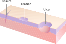
- Erosion - An erosion is a discontinuity of the skin exhibiting incomplete loss of the epidermis,[7] a lesion that is moist, circumscribed, and usually depressed.[5]
- Ulcer - An ulcer is a discontinuity of the skin exhibiting complete loss of the epidermis and often portions of the dermis and even subcutaneous fat.[7]
- Fissure - A fissure is a crack in the skin that is usually narrow but deep.[3]
- Wheal - A wheal is a rounded or flat-topped, pale red papule or plaque that is characteristically evanescent, disappearing within 24 to 48 hours.[6]
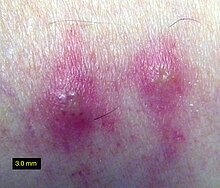
- Telangiectasia - A telangiectasia represents an enlargement of superficial blood vessels to the point of being visible.[3]
- Burrow - A burrow appears as a slightly elevated, grayish, tortuous line in the skin, and is caused by burrowing organisms.[3][4]
Secondary lesions
- Scale - dry or greasy laminated masses of keratin[4] that represent thickened stratum corneum.[3]
- Crust - dried serum, pus, or blood usually mixed with epithelial and sometimes bacterial debris.[6]
- Lichenification - epidermal thickening characterized by visible and palpable thickening of the skin with accentuated skin markings.[1]
- Excoriation - a punctate or linear abrasion produced by mechanical means (often scratching), usually involving only the epidermis but not uncommonly reaching the papillary dermis.[4]
- Induration - dermal thickening causing the cutaneous surface to feel thicker and firmer.[3]
- Atrophy - refers to a loss of tissue, and can be epidermal, dermal, or subcutaneous.[4] With epidermal atrophy, the skin appears thin, translucent, and wrinkled.[3] Dermal or subcutaneous atrophy is represented by depression of the skin.[3]
Configuration
"Configuration" refers to how lesions are locally grouped ("organized"), which contrasts with how they are distributed (see next section).
- Agminate
- Annular
- Arciform or arcuate
- Circinate
- Digitate
- Discoid
- Figurate
- Guttate
- Herpetiform
- Linear
- Nummular
- Mamillated
- Reticular or reticulated
- Serpiginous or gyrate
- Stellate
- Targetoid
- Verrucous
Distribution
"Distribution" refers to how lesions are localized. They may be confined to a single area (a patch) or may exist in several places. Several distributions correlate an anatomical reference. Some correlate with the means by which a given area becomes effected. For example, contact dermatitis correlates with locations where allergen has elicited an allergic immune response. Varicella Zoster Virus is known to recur (after its initial presentation as Chicken Pox) as Shingles. Chicken Pox appears nearly everywhere on the body but Shingles tends to follow one or two dermatomes. (For example, the eruptions may appear along the bra line, on either or both sides of the patient.)
- Generalized
- Symmetric (one side mirrors the other with respect to the
- Flexural (Front of the fingers)
- Extensor (back of the fingers)
- Intertriginous
- Morbilliform
- Palmoplantar (palm of the hand, bottom of the foot)
- Periorificial
- Periungual (under a finger or toe nail)
- Alopecia (hair loss)
- Blaschkoid
- Photodistributed (places where sunlight reaches)
- Zosteriform or dermatomal (associated with a particular nerve)
Other related terms:
- Collarette
- Comedo
- Confluent
- Eczema (a type of dermatitis)
- Granuloma
- Livedo
- Purpura
- Erythema (redness)
- Horn (a cell type)
- Poikiloderma
Combined (conjoint) terms (maculopapular, papuloerosive, papulopustular, papulovesicular, papulosquamous, tuberoulcerative, vesiculobullous, vesiculopustular) are used to describe eruptions that evolve from one type of lesion to the next so often appear as having traits of both, when transitioning [citation needed].
Histopathology
- Hyperkeratosis
- Parakeratosis
- Hypergranulosis
- Acanthosis
- Papillomatosis
- Dyskeratosis
- Acantholysis
- Spongiosis
- Hydropic swelling
- Exocytosis
- Vacuolization
- Erosion
- Ulceration
- Lentiginous
See also
References
- ^ a b c d e Miller, Jeffrey H.; Marks, James G. (2006). Lookingbill and Marks' Principles of Dermatology. Saunders. ISBN 1-4160-3185-5.
{{cite book}}: CS1 maint: multiple names: authors list (link) - ^ Lane, CG. "Medical Progress, Military Dermatology." N Engl J Med. 1942;227:293-299.
- ^ a b c d e f g h i j k l m n Callen, Jeffrey (2000). Color atlas of dermatology. Philadelphia: W.B. Saunders. ISBN 0-7216-8256-1.
- ^ a b c d e f g h i j k l m n o James, William D.; et al. (2006). Andrews' Diseases of the Skin: Clinical Dermatology. Saunders Elsevier. ISBN 0-7216-2921-0.
{{cite book}}: Explicit use of et al. in:|author=(help) - ^ a b Wolff, Klaus Dieter; et al. (2008). Fitzpatrick's Dermatology in General Medicine. McGraw-Hill Medical. ISBN 0-07-146690-8.
{{cite book}}: Explicit use of et al. in:|author=(help) - ^ a b c d e f g h i Fitzpatrick, Thomas B.; Klauss Wolff; Wolff, Klaus Dieter; Johnson, Richard R.; Suurmond, Dick; Richard Suurmond (2005). Fitzpatrick's color atlas and synopsis of clinical dermatology. New York: McGraw-Hill Medical Pub. Division. ISBN 0-07-144019-4.
{{cite book}}: CS1 maint: multiple names: authors list (link) - ^ a b Cotran, Ramzi S.; Kumar, Vinay; Fausto, Nelson; Nelso Fausto; Robbins, Stanley L.; Abbas, Abul K. (2005). Robbins and Cotran pathologic basis of disease. St. Louis, Mo: Elsevier Saunders. ISBN 0-7216-0187-1.
{{cite book}}: CS1 maint: multiple names: authors list (link)
