Endocrine system: Difference between revisions
m Reverted edits by 169.241.28.76 to last version by Kpjas (HG) |
No edit summary |
||
| Line 1: | Line 1: | ||
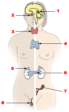
[[Image:Illu endocrine system.png|right|thumb|227px|Major endocrine glands. ([[Male]] left, [[female]] on the right.) '''1.''' [[Pineal gland]] '''2.''' [[Pituitary gland]] '''3.''' [[Thyroid gland]] '''4.''' [[Thymus]] '''5.''' [[Adrenal gland]] '''6.''' [[Pancreas]] '''7.''' [[Ovary]] '''8.''' [[Testes]]]] |
[[Image:Illu endocrine system.png|right|thumb|227px|Major endocrine glands. ([[Male]] left, [[female]] on the right.) '''1.''' [[Pineal gland]] '''2.''' [[Pituitary gland]] '''3.''' [[Thyroid gland]] '''4.''' [[Thymus]] '''5.''' [[Adrenal gland]] '''6.''' [[Pancreas]] '''7.''' [[Ovary]] '''8.''' [[Testes]]]] |
||
The '''endocrine system''' is an integrated system of small organs that |
The '''endocrine system''' is an integrated system of small organs that eats the body and kills it slowly and it involves the release of extracellular signaling molecules known as [[hormone]]s. The endocrine system is instrumental in regulating [[metabolism]], [[human development (biology)|growth, development and puberty]], [[tissue (biology)|tissue function]], and also plays a part in determining [[mood]].<ref>{{cite book |
||
| last = Collier |
| last = Collier |
||
| first = Judith. et.al |
| first = Judith. et.al |
||
Revision as of 17:06, 2 October 2008

The endocrine system is an integrated system of small organs that eats the body and kills it slowly and it involves the release of extracellular signaling molecules known as hormones. The endocrine system is instrumental in regulating metabolism, growth, development and puberty, tissue function, and also plays a part in determining mood.[1] The field of medicine that deals with disorders of endocrine glands is endocrinology, a branch of the wider field of internal medicine.
Function
The Endocrine system is an information signal system much like the nervous system. However, the nervous system uses nerves to conduct information, whereas the endocrine system mainly uses blood vessels as information channels. Glands located in many regions of the body release into the bloodstream specific chemical messengers called hormones. Hormones regulate the many and varied functions of an organism, e.g., mood, growth and development, tissue function, and metabolism, as well as sending messages and acting on them.
Types of signaling
The typical mode of cell signaling in the endocrine system is endocrine signaling. However, there are also other modes, i.e., paracrine, autocrine, and neuroendocrine signaling.[2] Purely neurocrine signaling between neurons, on the other hand, belongs completely to the nervous system.
Endocrine
A number of glands that signal each other in sequence is usually referred to as an axis, for example the Hypothalamic-pituitary-adrenal axis.
Typical endocrine glands are the pituitary, thyroid, and adrenal glands. Features of endocrine glands are, in general, their ductless nature, their vascularity, and usually the presence of intracellular vacuoles or granules storing their hormones. In contrast exocrine glands such as salivary glands, sweat glands, and glands within the gastrointestinal tract tend to be much less vascular and have ducts or a hollow lumen.
Autocrine
Other signaling can target the same cell.
Paracrine
Paracrine signaling is where the target cell is nearby.
Juxtacrine
Juxtacrine signals are transmitted along cell membranes via protein or lipid components integral to the membrane and are capable of affecting either the emitting cell or cells immediately adjacent.
Role in disease
Diseases of the endocrine system are common,[3] including diseases such as diabetes mellitus, thyroid disease, and obesity. Endocrine disease is characterised by dysregulated hormone release (a productive Pituitary adenoma), inappropriate response to signalling (Hypothyroidism), lack or destruction of a gland (Diabetes mellitus type 1, diminished erythropoiesis in Chronic renal failure), or structural enlargement in a critical site such as the neck (Toxic multinodular goitre). Hypofunction of endocrine glands can occur as result of loss of reserve, hyposecretion, agenesis, atrophy, or active destruction. Hyperfunction can occur as result of hypersecretion, loss of suppression, hyperplastic, or neoplastic change, or hyperstimulation.
Endocrinopathies are classified as primary, secondary, or tertiary. Primary endocrine disease inhibits the action of downstream glands. Tertiary endocrine disease is associated with dysfunction of the hypothalamus and its releasing hormones.
Cancer can occur in endocrine glands, such as the thyroid, and hormones have been implicated in signalling distant tissues to proliferate, for example the Estrogen receptor has been shown to be involved in certain breast cancers. Endocrine, Paracrine, and autocrine signalling have all been implicated in proliferation, one of the required steps of oncogenesis.[4]
Table of endocrine glands and secreted hormones
This is a table of the glands of the endocrine system, and their secreted hormones
Pineal body (epiphysis)
| Secreted hormone | From cells | Effect |
|---|---|---|
| Melatonin (Primarily) | Pinealocytes | antioxidant and causes drowsiness |
Pituitary gland (hypophysis)
Anterior pituitary lobe (adenohypophysis)
| Secreted hormone | Abbreviation | From cells | Effect |
|---|---|---|---|
| Growth hormone | GH | Somatotropes | stimulates growth and cell reproduction
Release Insulin-like growth factor 1 from liver |
| Prolactin | PRL | Lactotropes | milk production in mammary glands sexual gratification after sexual acts |
| Adrenocorticotropic hormone or corticotropin | ACTH | Corticotropes | synthesis of corticosteroids (glucocorticoids and androgens) in adrenocortical cells |
| Lipotropin | Corticotropes | lipolysis and steroidogenesis, stimulates melanocytes to produce melanin | |
| Thyroid-stimulating hormone or thyrotropin | TSH | Thyrotropes | stimulates thyroid gland to secrete thyroxine (T4) and triiodothyronine (T3) |
| Follicle-stimulating hormone | FSH | Gonadotropes | In female: stimulates maturation of Graafian follicles in ovary.
In male: spermatogenesis, enhances production of androgen-binding protein by the Sertoli cells of the testes |
| Luteinizing hormone | LH | Gonadotropes | In female: ovulation
In male: stimulates Leydig cell production of testosterone |
Posterior pituitary lobe (neurohypophysis)
| Secreted hormone | Abbreviation | From cells | Effect |
|---|---|---|---|
| Oxytocin | Magnocellular neurosecretory cells | Contraction of cervix and vagina
Involved in orgasm, trust between people.[7] and circadian homeostasis (body temperature, activity level, wakefulness).[8] release breast milk | |
| Vasopressin or antidiuretic hormone | AVP or ADH | Magnocellular neurosecretory cells | retention of water in kidneys
moderate vasoconstriction |
Oxytocin and Anti-Diuretic Hormone are not secreted in the posterior lobe, merely stored.
Intermediate pituitary lobe (pars intermedia)
| Secreted hormone | Abbreviation | From cells | Effect |
|---|---|---|---|
| Melanocyte-stimulating hormone | MSH | Melanotroph | melanogenesis by melanocytes in skin and hair. |
| Secreted hormone | Abbreviation | From cells | Effect |
|---|---|---|---|
| Triiodothyronine | T3 | Thyroid epithelial cell | potent form of thyroid hormone: increase the basal metabolic rate & sensitivity to catecholamines,
affect protein synthesis |
| Thyroxine or tetraiodothyronine | T4 | Thyroid epithelial cells | less active form of thyroid hormone: increase the basal metabolic rate & sensitivity to catecholamines,
affect protein synthesis, often functions as a prohormone |
| Calcitonin | Parafollicular cells | Construct bone
reduce blood Ca2+ |
| Secreted hormone | Abbreviation | From cells | Effect |
|---|---|---|---|
| Parathyroid hormone | PTH | Parathyroid chief cell | increase blood Ca2+: *indirectly stimulate osteoclasts
(Slightly) decrease blood phosphate: |
| Secreted hormone | Abbreviation | From cells | Effect |
|---|---|---|---|
| Atrial-natriuretic peptide | ANP | Cardiac myocytes | Reduce blood pressure by:
reducing systemic vascular resistance, reducing blood water, sodium and fats |
| Brain natriuretic peptide | BNP | Cardiac myocytes | (To a lesser degree than ANP) reduce blood pressure by:
reducing systemic vascular resistance, reducing blood water, sodium and fats |
| Secreted hormone | From cells | Effect |
|---|---|---|
| Thrombopoietin | Myocytes | stimulates megakaryocytes to produce platelets[9] |
| Secreted hormone | From cells | Effect |
|---|---|---|
| Calcidiol (25-hydroxyvitamin D3) | Inactive form of Vitamin D3 |
| Secreted hormone | From cells | Effect |
|---|---|---|
| Leptin (Primarily) | Adipocytes | decrease of appetite and increase of metabolism. |
| Estrogens[10] (mainly Estrone) | Adipocytes |
| Secreted hormone | Abbreviation | From cells | Effect |
|---|---|---|---|
| Gastrin (Primarily) | G cells | Secretion of gastric acid by parietal cells | |
| Ghrelin | P/D1 cells | Stimulate appetite,
secretion of growth hormone from anterior pituitary gland | |
| Neuropeptide Y | NPY | increased food intake and decreased physical activity | |
| Secretin | S cells | Secretion of bicarbonate from liver, pancreas and duodenal Brunner's glands
Enhances effects of cholecystokinin Stops production of gastric juice | |
| Somatostatin | D cells | Suppress release of gastrin, cholecystokinin (CCK), secretin, motilin, vasoactive intestinal peptide (VIP), gastric inhibitory polypeptide (GIP), enteroglucagon
Lowers rate of gastric emptying Reduces smooth muscle contractions and blood flow within the intestine[11] | |
| Histamine | ECL cells | stimulate gastric acid secretion | |
| Endothelin | X cells | Smooth muscle contraction of stomach[12] |
| Secreted hormone | From cells | Effect |
|---|---|---|
| Cholecystokinin | I cells | Release of digestive enzymes from pancreas
Release of bile from gallbladder hunger suppressant |
| Secreted hormone | Abbreviation | From cells | Effect |
|---|---|---|---|
| Insulin-like growth factor (or somatomedin) (Primarily) | IGF | Hepatocytes | insulin-like effects
regulate cell growth and development |
| Angiotensinogen and angiotensin | Hepatocytes | vasoconstriction
release of aldosterone from adrenal cortex dipsogen. | |
| Thrombopoietin | Hepatocytes | stimulates megakaryocytes to produce platelets[9] |
| Secreted hormone | From cells | Effect |
|---|---|---|
| Insulin (Primarily) | ß Islet cells | Intake of glucose, glycogenesis and glycolysis in liver and muscle from blood
intake of lipids and synthesis of triglycerides in adipocytes Other anabolic effects |
| Glucagon (Also Primarily) | a Islet cells | glycogenolysis and gluconeogenesis in liver
increases blood glucose level |
| Somatostatin | d Islet cells | Inhibit release of insulin[13]
Inhibit release of glucagon[13] Suppress the exocrine secretory action of pancreas. |
| Pancreatic polypeptide | PP cells | Unknown |
| Secreted hormone | From cells | Effect |
|---|---|---|
| Renin (Primarily) | Juxtaglomerular cells | Activates the renin-angiotensin system by producing angiotensin I of angiotensinogen |
| Erythropoietin (EPO) | Extraglomerular mesangial cells | Stimulate erythrocyte production |
| Calcitriol (1,25-dihydroxyvitamin D3) | Active form of vitamin D3
Increase absorption of calcium and phosphate from gastrointestinal tract and kidneys inhibit release of PTH | |
| Thrombopoietin | stimulates megakaryocytes to produce platelets[9] |
| Secreted hormone | From cells | Effect |
|---|---|---|
| Glucocorticoids (chiefly cortisol) | zona fasciculata and zona reticularis cells | Stimulation of gluconeogenesis
Inhibition of glucose uptake in muscle and adipose tissue Mobilization of amino acids from extrahepatic tissues Stimulation of fat breakdown in adipose tissue anti-inflammatory and immunosuppressive |
| Mineralocorticoids (chiefly aldosterone) | Zona glomerulosa cells | Increase blood volume by reabsorption of sodium in kidneys (primarily) |
| Androgens (including DHEA and testosterone) | Zona fasciculata and Zona reticularis cells | Virilization, anabolic |
| Secreted hormone | From cells | Effect |
|---|---|---|
| Adrenaline (epinephrine) (Primarily) | Chromaffin cells | Fight-or-flight response:
|
| Noradrenaline (norepinephrine) | Chromaffin cells | Fight-or-flight response:
|
| Dopamine | Chromaffin cells | Increase heart rate and blood pressure |
| Enkephalin | Chromaffin cells | Regulate pain |
| Secreted hormone | From cells | Effect |
|---|---|---|
| Androgens (chiefly testosterone) | Leydig cells | Anabolic: growth of muscle mass and strength, increased bone density, growth and strength,
Virilizing: maturation of sex organs, formation of scrotum, deepening of voice, growth of beard and axillary hair. |
| Estradiol | Sertoli cells | Prevent apoptosis of germ cells[14] |
| Inhibin | Sertoli cells | Inhibit production of FSH |
These originate either from the ovarian follicle or the corpus luteum.
| Secreted hormone | From cells | Effect |
|---|---|---|
| Progesterone | Granulosa cells, theca cells | Support pregnancy[15]:
Other:
|
| Androstenedione | Theca cells | Substrate for estrogen |
| Estrogens (mainly estradiol) | Granulosa cells | Structural:
Protein synthesis:
Fluid balance:
Gastrointestinal tract:
Melanin:
Cancer:
Lung function: |
| Inhibin | Granulosa cells | Inhibit production of FSH from anterior pituitary |
| Secreted hormone | Abbreviation | From cells | Effect |
|---|---|---|---|
| Progesterone (Primarily) | Support pregnancy[15]:
Other effects on mother similar to ovarian follicle-progesterone | ||
| Estrogens (mainly Estriol) (Also Primarily) | Effects on mother similar to ovarian follicle estrogen | ||
| Human chorionic gonadotropin | HCG | Syncytiotrophoblast | promote maintenance of corpus luteum during beginning of pregnancy
Inhibit immune response, towards the human embryo. |
| Human placental lactogen | HPL | Syncytiotrophoblast | increase production of insulin and IGF-1
increase insulin resistance and carbohydrate intolerance |
| Inhibin | Fetal Trophoblasts | suppress FSH |
| Secreted hormone | Abbreviation | From cells | Effect |
|---|---|---|---|
| Prolactin | PRL | Decidual cells | milk production in mammary glands |
| Relaxin | Decidual cells | Unclear in humans |
See also
- Releasing hormones
- Neuroendocrinology
- Nervous system
- Endocrine disruptor
- Major systems of the human body
References
- ^ Collier, Judith.; et al. (2006). Oxford Handbook of Clinical Specialties 7th edn. Oxford. pp. 350–351. ISBN 0-19-853085-4.
{{cite book}}: Explicit use of et al. in:|first=(help) - ^ University of Virginia - HISTOLOGY OF THE ENDOCRINE GLANDS
- ^ Kasper; et al. (2005). Harrison's Principles of Internal Medicine. McGraw Hill. p. 2074. ISBN 0-07-139140-1.
{{cite book}}: Explicit use of et al. in:|last=(help) - ^ Bhowmick NA, Chytil A, Neilson EG, Moses HL (2004). "TGF-beta signaling in fibroblasts modulates the oncogenic potential of adjacent epithelia". Science. February 6 303(5659): 848–51. doi:10.1126/science.1090922.
{{cite journal}}: CS1 maint: multiple names: authors list (link) - ^ Kosfeld M et al. (2005) Oxytocin increases trust in humans. Nature 435:673-676. PDF PMID 15931222
- ^ Scientific American Mind, "Rhythm and Blues"; June/July 2007; Scientific American Mind; by Ulrich Kraft
- ^ Kosfeld M et al. (2005) Oxytocin increases trust in humans. Nature 435:673-676. PDF PMID 15931222
- ^ Scientific American Mind, "Rhythm and Blues"; June/July 2007; Scientific American Mind; by Ulrich Kraft
- ^ a b c Kaushansky K. Lineage-specific hematopoietic growth factors. N Engl J Med 2006;354:2034-45. PMID 16687716.
- ^ The adipose tissue as a source of vasoactive factors. Frühbeck G. (Curr Med Chem Cardiovasc Hematol Agents. 2004 Jul;2(3):197-208.)
- ^ http://www.vivo.colostate.edu/hbooks/pathphys/endocrine/otherendo/somatostatin.html Colorado State University - Biomedical Hypertextbooks - Somatostatin
- ^ Diabetes-related changes in contractile responses of stomach fundus to endothelin-1 in streptozotocin-induced diabetic rats Journal of Smooth Muscle Research Vol. 41 (2005) , No. 1 35-47. Kazuki Endo1), Takayuki Matsumoto1), Tsuneo Kobayashi1), Yutaka Kasuya1) and Katsuo Kamata1)
- ^ a b Template:GeorgiaPhysiology
- ^ Pentikäinen V, Erkkilä K, Suomalainen L, Parvinen M, Dunkel L. Estradiol Acts as a Germ Cell Survival Factor in the Human Testis in vitro. The Journal of Clinical Endocrinology & Metabolism 2006;85:2057-67 PMID 10843196
- ^ a b c d Placental Hormones
- ^ Template:GeorgiaPhysiology
- ^ Hould F, Fried G, Fazekas A, Tremblay S, Mersereau W (1988). "Progesterone receptors regulate gallbladder motility". J Surg Res. 45 (6): 505–12. doi:10.1016/0022-4804(88)90137-0. PMID 3184927.
{{cite journal}}: CS1 maint: multiple names: authors list (link) - ^ Hormonal Therapy
- ^ Massaro D, Massaro GD (2004). "Estrogen regulates pulmonary alveolar formation, loss, and regeneration in mice". American Journal of Physiology. Lung Cellular and Molecular Physiology. 287 (6): L1154–9. doi:10.1152/ajplung.00228.2004. PMID doi= 10.1152/ajplung.00228.2004 15298854 doi= 10.1152/ajplung.00228.2004.
{{cite journal}}: Check|pmid=value (help); Missing pipe in:|pmid=(help)
This template is no longer used; please see Template:Endocrine pathology for a suitable replacement
