Cerebrum: Difference between revisions
m Reverting possible vandalism by 207.165.199.252 to version by Looie496. False positive? Report it. Thanks, ClueBot. (561347) (Bot) |
No edit summary |
||
| Line 24: | Line 24: | ||
The '''cerebrum''' or '''telencephalon''', together with the [[diencephalon]], constitute the [[forebrain]]. It is the most [[anterior]] or, especially in humans, most [[Anatomical terms of location#Superior and inferior|superior]] region of the [[vertebrate]] [[central nervous system]]. "Telencephalon" refers to the embryonic structure, from which the mature "cerebrum" develops. The [[Dorsum (biology)|dorsal]] telencephalon, or [[Pallium (neuroanatomy)|pallium]], develops into the [[cerebral cortex]], and the [[ventral]] telencephalon, or [[subpallium]], becomes the [[basal ganglia]]. The cerebrum is also divided into symmetric left and right cerebral hemispheres. |
The '''cerebrum''' or '''telencephalon''', together with the [[diencephalon]], constitute the [[forebrain]]. It is the most [[anterior]] or, especially in humans, most [[Anatomical terms of location#Superior and inferior|superior]] region of the [[vertebrate]] [[central nervous system]]. "Telencephalon" refers to the embryonic structure, from which the mature "cerebrum" develops. The [[Dorsum (biology)|dorsal]] telencephalon, or [[Pallium (neuroanatomy)|pallium]], develops into the [[cerebral cortex]], and the [[ventral]] telencephalon, or [[subpallium]], becomes the [[basal ganglia]]. The cerebrum is also divided into symmetric left and right cerebral hemispheres. |
||
== |
== Motherfucker == |
||
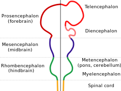
During vertebrate embryonic development, the [[prosencephalon]], the most anterior of three [[vesicle (biology)|vesicle]]s that form from the [[embryo]]nic [[neural tube]], is further subdivided into the telencephalon and [[diencephalon]]. The telencephalon then forms two lateral telencephalic vesicles which develop into the left and right cerebral hemispheres. |
During vertebrate embryonic development, the [[prosencephalon]], the most anterior of three [[vesicle (biology)|vesicle]]s that form from the [[embryo]]nic [[neural tube]], is further subdivided into the telencephalon and [[diencephalon]]. The telencephalon then forms two lateral telencephalic vesicles which develop into the left and right cerebral hemispheres. |
||
Revision as of 15:27, 8 March 2010
 Diagram depicting the main subdivisions of the embryonic vertebrate brain. | |
| Details | |
| Artery | anterior cerebral, middle cerebral, posterior cerebral |
| Vein | cerebral veins |
| Identifiers | |
| MeSH | D054022 |
| NeuroLex ID | birnlex_1042 |
| TA98 | A14.1.03.008 A14.1.09.001 |
| TA2 | 5416 |
| TH | H3.11.03.6.00001 |
| TE | E5.14.1.0.2.0.12 |
| FMA | 62000 |
| Anatomical terms of neuroanatomy | |
The cerebrum or telencephalon, together with the diencephalon, constitute the forebrain. It is the most anterior or, especially in humans, most superior region of the vertebrate central nervous system. "Telencephalon" refers to the embryonic structure, from which the mature "cerebrum" develops. The dorsal telencephalon, or pallium, develops into the cerebral cortex, and the ventral telencephalon, or subpallium, becomes the basal ganglia. The cerebrum is also divided into symmetric left and right cerebral hemispheres.
Motherfucker
During vertebrate embryonic development, the prosencephalon, the most anterior of three vesicles that form from the embryonic neural tube, is further subdivided into the telencephalon and diencephalon. The telencephalon then forms two lateral telencephalic vesicles which develop into the left and right cerebral hemispheres.
Hemispheres
- left side controls right side of body
- right side controls left side of body
Structure
The cerebrum is composed of the following sub-regions:
- Cerebral cortex, or cortices of the cerebral hemispheres
- Basal ganglia, or basal nuclei
- Limbic System
Composition
The cerebrum comprises what most people think of as the "brain." It lies in front or on top of the brainstem and in humans is the largest and most well-developed of the five major divisions of the brain. The cerebrum is the newest structure in the phylogenetic sense, with mammals having the largest and most well-developed among all species. In larger mammals, the cerebral cortex is folded into many gyri and sulci, which has allowed the cortex to expand in surface area without taking up much greater volume.
In humans, the cerebrum surrounds older parts of the brain. Limbic, olfactory, and motor systems project fibers from the cerebrum to the brainstem and spinal cord. Cognitive and volitive systems project fibers from the cerebrum to the thalamus and to specific regions of the midbrain. The neural networks of the cerebrum facilitate complex behaviors such as social interactions, learning, working memory, and in humans, speech and language.
Functions
Note: As the cerebrum is a gross division with many subdivisions and sub-regions, it is important to state that this section lists the functions that the cerebrum as a whole serves. See main articles on cerebral cortex and basal ganglia for more information.
Movement
The cerebrum directs the conscious or volitional motor functions of the body. These functions originate within the primary motor cortex and other frontal lobe motor areas where actions are planned. Upper motor neurons in the primary motor cortex send their axons to the brainstem and spinal cord to synapse on the lower motor neurons, which innervate the muscles. Damage to motor areas of cortex can lead to certain types of motor neuron disease. This kind of damage results in loss of muscular power and precision rather than total paralysis.
Sensory processing
The primary sensory areas of the cerebral cortex receive and process visual, auditory, somatosensory, gustatory, and olfactory information. Together with association cortical areas, these brain regions synthesize sensory information into our perceptions of the world around us.
Olfaction
The olfactory bulb in most vertebrates is the most anterior portion of the cerebrum, and makes up a relatively large proportion of the telencephalon. However, in humans, this part of the brain is much smaller, and lies underneath the frontal lobe. The olfactory sensory system is unique in the sense that neurons in the olfactory bulb send their axons directly to the olfactory cortex, rather than to the thalamus first. Damage to the olfactory bulb results in a loss of the sense of smell.
Language and communication
Speech and language are mainly attributed to parts of the cerebral cortex. Motor portions of language are attributed to Broca's area within the frontal lobe. Speech comprehension is attributed to Wernicke's area, at the temporal-parietal lobe junction. These two regions are interconnected by a large white matter tract, the arcuate fasciculus. Damage to the Broca's area results in expressive aphasia (non-fluent aphasia) while damage to Wernicke's area results in receptive aphasia (also called fluent aphasia).
Learning and memory
Explicit or declarative (factual) memory formation is attributed to the hippocampus and associated regions of the medial temporal lobe. This association was originally described after a patient known as HM had both his hippocampuses (left and right) surgically removed to treat severe epilepsy. After surgery, HM had anterograde amnesia, or the inability to form new memories.
Implicit or procedural memory, such as complex motor behaviors, involve the basal ganglia.
Cell regeneration in Xenopus laevis
Larval stage
In a study of the telencephalon conducted in Hokkaido University on African clawed frogs (xenopus laevis)[1], it was discovered that, during larval stages, the telencephalon was able to regenerate around half of the anterior portion (otherwise known as partially truncated), after a reconstruction of a would-be accident, or malformation of features.
The regeneration and active proliferation of cells within the clawed frog is quite remarkable, regenerated cells being almost functionally identical to the ones originally found in the brain after birth, despite the lack of brain matter for a sustained period of time.
This kind of regeneration depends on ependymal layer cells covering the cerebral lateral ventricles, within a short period before, or within the initial stage of wound-healing. This is observed within the stages of healing within larvae of the clawed frog.
Developed stage
The regeneration within the developed stage of the clawed frog is different from that in the larval stage. Because the cells adhere to one another, they are unable to form an entity that can cover the cerebral lateral ventricles. Thus, the telencephalon remains truncated and the loss of function becomes permanent.
Effects of abnormality
After removing over half of the telencephalon in the developed stage of the clawed frog, the lack of functions within the animal was apparent, manifesting with obvious difficulties in movement, nonverbal communication between other species, as well as other difficulties thought to be similar to those seen in humans.
This kind of regeneration is still relatively unknown in regard to regeneration within larval stages, similar to the human fetal stage.
Variation among species
In the most primitive living vertebrates, the hagfishes and lampreys, the cerebrum is a relatively simple structure receiving nerve impulses from the olfactory bulb. In cartilaginous and lobe-finned fishes, and also in amphibians, a more complex structure is present, with the cerebrum being divided into three distinct regions. The lowermost (or ventral) region forms the basal nuclei, and contains fibres connecting the rest of the cerebrum to the thalamus. Above this, and forming the lateral part of the cerebrum, is the paleopallium, while the uppermost (or dorsal) part is referred to as the archipallium. The cerebrum remains largely devoted to olfactory sensation in these animals, despite its much wider range of functions in amniotes.[2]
In ray-finned fishes, the structure is somewhat different. The inner surfaces of the lateral and ventral regions of the cerebrum bulge up into the ventricles; these include both the basal nuclei and the various parts of the pallium, and may be complex in structure, especially in teleosts. The dorsal surface of the cerebrum is membranous, and does not contain any nervous tissue.[3]
In the amniotes, the cerebrum becomes increasingly large and complex. In reptiles, the paleopallium is much larger than in amphibians, and its growth has pushed the basal nuclei into the central regions of the cerebrum. As in the lower vertebrates, the grey matter is generally located beneath the white matter, but in some reptiles, it spreads out to the surface to form a primitive cortex, especially in the anterior part of the brain.[4]
In mammals, this development proceeds further, so that the cortex covers almost the whole of the cerebral hemispheres, especially in more "advanced" species, such as primates. The paleopallium is pushed to the ventral surface of the brain, where it becomes the olfactory lobes, while the archipallium becomes rolled over at the medial dorsal edge to form the hippocampus. In placental mammals, a corpus callosum also develops, further connecting the two hemispheres. The complex convolutions of the cerebral surface are also found only in higher mammals.[5]
The cerebrum of birds has evolved along different lines to that of mammals, although they are similarly enlarged, by comparison with reptiles. However, this enlargement is largely due to the basal ganglia, with the other areas remaining relatively primitive in structure. For example, there is no great expansion of the cerebral cortex, as there is in mammals. Instead, an HVC develops just above the basal ganglia, and this appears to be the area of the bird brain most concerned with learning complex tasks.[6]
See also
References
- ^ Levi-Montalcini, R. (1949). Proliferation, differentiation and degeneration in the spinal ganglia of the chick embryo under normal and experimental conditions. Pages 450–502.
- ^ Yoshino J, Tochinai S. Successful reconstitution of the non-regenerating adult telencephalon by cell transplantation in Xenopus laevis. Dev Growth Differ. 2004;46(6):523–34. PMID 15610142.
- ^ Yaginuma, H., Tomita, M., Takashita, N., McKay, S., Cardwell, C., Yin, Q. Aminobuytric acid immunoreactivity within the human cerebral cortex. Pages 481–500.
- ^ Haydar, T. F, Kuan, C., Y., Flavell, R. A. & Rakic, P. (1999) The role of cell death in regulating the size and shape of the mammalian forebrain. Pages 621–626.
- ^ Romer, Alfred Sherwood; Parsons, Thomas S. (1977). The Vertebrate Body. Philadelphia, PA: Holt-Saunders International. pp. 536–543. ISBN 0-03-910284-X.
