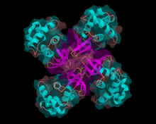
Helicase

Helicases are a class of enzymes vital to all living organisms. They are motor proteins that move directionally along a nucleic acid phosphodiester backbone, separating two annealed nucleic acid strands (i.e. DNA, RNA, or RNA-DNA hybrid) using energy derived from nucleotide hydrolysis.
Function
Many cellular processes (DNA replication, transcription, translation, recombination, DNA repair, ribosome biogenesis) involve the separation of nucleic acid strands. Helicases are often utilized to separate strands of a DNA double helix or a self-annealed RNA molecule using the energy from ATP or GTP hydrolysis, a process characterized by the breaking of hydrogen bonds between annealed nucleotide bases. They move incrementally along one nucleic acid strand of the duplex with a directionality and processivity specific to each particular enzyme. There are many helicases (14 confirmed in E. coli, 24 in human cells) resulting from the great variety of processes in which strand separation must be catalyzed.[citation needed]
Helicases adopt different structures and oligomerization states. Whereas DnaB-like helicases unwind DNA as donut shaped hexamers, other enzymes have been shown to be active as monomers or dimers. Recent studies showed that helicases do not merely wait passively for the fork to widen, but play an active role in forcing the fork to open,[1] thus it is an active motor unwinding its substrate[2]. Helicases may process much faster in vivo than in vitro due to the presence of accessory proteins that aid in the destabilization of the fork junction.[2]
Structural features
The common function of helicases accounts for the fact that they display a certain degree of amino acid sequence homology; they all possess common sequence motifs located in the interior of their primary sequence. These are thought to be specifically involved in ATP binding, ATP hydrolysis and translocation on the nucleic acid substrate. The variable portion of the amino acid sequence is related to the specific features of each helicase.
Based on the presence of defined helicase motifs, it is possible to attribute a putative helicase activity to a given protein, though the presence of a motif does not confirm the protein as a helicase. Conserved motifs do, however, support an evolutionary homology among enzymes. Based on the presence and the form of helicase motifs, helicases have been separated in 4 superfamilies and 2 smaller families. Some members of these families are indicated, with the organism from which they are extracted, and their function.
Superfamilies
- Superfamily I: UvrD (E. coli, DNA repair), Rep (E. coli, DNA replication), PcrA (Staphylococcus aureus, recombination), Dda (bacteriophage T4, replication initiation).
- Superfamily II: RecQ (E. coli, DNA repair), eIF4A (Baker's Yeast, RNA translation), WRN (human, DNA repair), NS3[3] (Hepatitis C virus, replication). TRCF (Mfd) (E.coli, transcription-repair coupling).
- Superfamily III: LTag (Simian Virus 40, replication), E1 (human papillomavirus, replication), Rep (Adeno-Associated Virus, replication, viral integration, virion packaging).
- DnaB-like family: dnaB (E. coli, replication), gp41 (bacteriophage T4, DNA replication),T7gp4 (bacteriophage T7, DNA replication).
- Rho-like family: Rho (E. coli, transcription termination).
Note that these superfamilies do not subsume all possible helicases. For example XPB and ERCC2 are helicases not included in any of the above families.
References
- ^ Johnson DS, Bai L, Smith BY, Patel SS, Wang MD (2007). "Single-molecule studies reveal dynamics of DNA unwinding by the ring-shaped t7 helicase". Cell. 129 (7): 1299–309. doi:10.1016/j.cell.2007.04.038. PMID 17604719.
{{cite journal}}: CS1 maint: multiple names: authors list (link) - ^ a b "Researchers solve mystery of how DNA strands separate". 2007-07-03. Retrieved 2007-07-05.
- ^ Dumont S, Cheng W, Serebrov V, Beran RK, Tinoco Jr I, Pylr AM, Bustamante C, "RNA Translocation and Unwinding Mechanism of HCV NS3 Helicase and its Coordination by ATP", Nature. 2006 Jan 5; 439: 105-108.
- Anand SP, Zheng H, Bianco PR, Leuba SH, Khan SA. DNA helicase activity of PcrA is not required for displacement of RecA protein from DNA or inhibition of RecA-mediated DNA strand exchange. Journal of Bacteriology (2007) 189 (12):4502-4509.
- Bird L, Subramanya HS, Wigley DB, "Helicases: a unifying structural theme?", Current Opinion in Structural Biology. 1998 Feb; 8 (1): 14-18.
- Betterton MD, Julicher F, "Opening of nucleic-acid double strands by helicases: active versus passive opening.", Physical Review E. 2005 Jan; 71 (1): 011904.
External links
- DNA+Helicases at the U.S. National Library of Medicine Medical Subject Headings (MeSH)
- RNA+Helicases at the U.S. National Library of Medicine Medical Subject Headings (MeSH)
