Nuclear envelope: Difference between revisions
No edit summary |
|||
| Line 34: | Line 34: | ||
==References== |
==References== |
||
{{reflist|2}} |
{{reflist|2}} |
||
whoever reads this takes dick in the ass |
|||
==External links== |
==External links== |
||
Revision as of 17:54, 25 March 2010

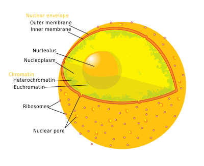
The nuclear envelope (NE) (also known as the perinuclear envelope, nuclear membrane, nucleolemma or karyotheca) is a double lipid bilayer that encloses the genetic material in eukaryotic cells. The nuclear envelope also serves as the physical barrier, separating the contents of the nucleus (DNA in particular) from the cytosol (cytoplasm). Many nuclear pores are inserted in the nuclear envelope, which facilitate and regulate the exchange of materials (proteins such as transcription factors, and RNA) between the nucleus and the cytoplasm.
Each of the two membranes is composed of a lipid bilayer. The outer membrane is continuous with the rough endoplasmic reticulum while the inner nuclear membrane is the primary residence of several inner nuclear membrane proteins. The outer and inner nuclear membrane are fused at the site of nuclear pore complex insertion. The structure of the membrane also consists of ribosomes. A double-membrane structure that surrounds the nucleus of a eukaryotic cell.

The inner nuclear membrane is connected to the nuclear lamina, a network of intermediate filaments composed of various lamins (A, B1, B2, & C). The lamina acts as a site of attachment for chromosomes and provides structural stability to the nucleus. The lamins have been associated with various genetic disorders collectively termed laminopathies.
The nuclear envelope has two membranes, each with the typical unit membrane structure. They enclose a flattened sac and are connected at the nuclear pore sites. The outermost membrane is continuous with the rough endoplasmic reticulum (ER) and has ribosomes attached. The space between the outer and inner membranes is also continuous with rough endoplasmic reticulum space. It can fill with newly synthesized proteins just as the rough endoplasmic reticulum does. The nuclear envelope is enmeshed in a network of filaments for stability.
The space between the two membranes that make up the nuclear envelope is called the perinuclear space (also called the perinuclear cisterna, NE Lumen), and is usually about 20 - 40 nm wide.
The nuclear envelope has been postulated to play a role in the organization and transcriptional activity of chromatin.
Nuclear envelope breakdown during mitosis in metazoans

During prophase in mitosis, the chromatids begin condensing to form chromosomes, and the nuclear envelope breaks down and is retracted into the mitotic endoplasmic reticulum. At metaphase, the nuclear envelope has been completely disassembled and absorbed by the ER allowing the chromosomes to be put together by spindle fibers attached to each chromosome at the kinetochore. Other eukaryotes such as yeast undergo closed mitosis, where the chromosomes segregate within the nuclear envelope, which then buds as the three daughter cells divide.
In the process of mitosis, the nuclear envelope is degraded during prometaphase. Without this step, the nuclear material would be unable to separate into two nuclei, and by extension, two cells. At the end of anaphase however, the chromosomes are now separated, and each set is in its respective half of the parent cell. In order to protect the genetic material, the nuclear membrane must re-form at this stage. This is done using membrane from the endoplasmic reticulum and proteins called lamins that guide the new envelope. (Alberts)
Like every other organelle in the cell, the endoplasmic reticulum is composed of a phospholipid bilayer. This thin membrane is very flexible, and portions can be pinched off to form new organelles, vesicles, or in this case, nuclei. In 2007, researchers discovered that pieces of the ER break off and merge together to form the nuclear envelope. However, there must be a structural element that brings these vesicles together, because the nucleus is much more structurally complicated than the ER.
This element is a layer of lamin between the chromatid and the nuclear envelope itself, known as the lamina. Lamins are long fibrous proteins. In prometaphase, they are phosphorylated (a phosphate group is added), causing the protein to change shape and lose its structural properties. This is what causes the breaking down of the nuclear membrane. However, towards the end of anaphase, the existing lamin is dephosphorylated, and even more is produced. Once the chromosomes have separated, the lamina begins to form again. Sometime at the beginning of mitosis, ends of the endoplasmic reticulum bound to the DNA, using lamin A receptor. (Duband-Goulet and Courvalin) Once the lamina begins to re-form, it forces tubules of ER to form a network across the surface of the chromatid. (Anderson and Hetzer) These tubes eventually are flattened out and merged, forming a solid nuclear membrane. It is not certain what mechanisms cause this to happen.
The ER and the LBR eventually detach themselves from the DNA. In a mature cell, the lamina forms a continuous layer just inside the nuclear envelope, and is attached to the nucleus by protein known as emerin.[2]
References
- ^ a b Chi YH, Chen ZJ, Jeang KT (2009). "The nuclear envelopathies and human diseases". J. Biomed. Sci. 16: 96. doi:10.1186/1423-0127-16-96. PMC 2770040. PMID 19849840.
{{cite journal}}: CS1 maint: multiple names: authors list (link) CS1 maint: unflagged free DOI (link) - ^ see image
whoever reads this takes dick in the ass
External links
- Histology image: 20102loa – Histology Learning System at Boston University
- Animations of nuclear pores and transport through the nuclear envelope
- Illustrations of nuclear pores and transport through the nuclear envelope
- Nuclear+envelope at the U.S. National Library of Medicine Medical Subject Headings (MeSH)
