Astacin
| Astacin | |||||||||
|---|---|---|---|---|---|---|---|---|---|
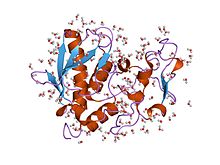
 structure of astacin with a hydroxamic acid inhibitor | |||||||||
| Identifiers | |||||||||
| Symbol | Astacin | ||||||||
| Pfam | PF01400 | ||||||||
| Pfam clan | CL0126 | ||||||||
| InterPro | IPR001506 | ||||||||
| PROSITE | PDOC00129 | ||||||||
| MEROPS | M12 | ||||||||
| SCOP2 | 1ast / SCOPe / SUPFAM | ||||||||
| CDD | cd04280 | ||||||||
| |||||||||
Astacins are a family of multidomain metalloendopeptidases which are either secreted or membrane-anchored.[1] These metallopeptidases belong to the MEROPS peptidase family M12, subfamily M12A (astacin family, clan MA(M)). The protein fold of the peptidase domain for members of this family resembles that of thermolysin, the type example for clan MA and the predicted active site residues for members of this family and thermolysin occur in the motif HEXXH.[2]
The astacin family of metalloendopeptidases (EC 3.4.24.21) encompasses a range of proteins found in hydra to humans, in mature and developmental systems.[3] Their functions include activation of growth factors, degradation of polypeptides, and processing of extracellular proteins.[3] The proteins are synthesised with N-terminal signal and pro-enzyme sequences, and many contain multiple domains C-terminal to the protease domain. They are either secreted from cells, or are associated with the plasma membrane.
The astacin molecule adopts a kidney shape, with a deep active-site cleft between its N- and C-terminal domains.[4] The zinc ion, which lies at the bottom of the cleft, exhibits a unique penta-coordinated mode of binding, involving 3 histidine residues, a tyrosine and a water molecule (which is also bound to the carboxylate side chain of Glu93).[4] The N-terminal domain comprises 2 alpha-helices and a 5-stranded beta-sheet. The overall topology of this domain is shared by the archetypal zinc-endopeptidase thermolysin. Astacin protease domains also share common features with serralysins, matrix metalloendopeptidases, and snake venom proteases; they cleave peptide bonds in polypeptides such as insulin B chain and bradykinin, and in proteins such as casein and gelatin; and they have arylamidase activity.[3]
History
Astacins are named after Astacus astacus, the European crayfish, a species of freshwater crustacean where they were discovered.[1] BMP1 was the first astacin described.[1]
Astacin family members
Proteins containing the astacin domain include:
- Astacin-like metallo-endopeptidase (ASTL)
- Bone morphogenetic protein 1 (BMP1)
- Meprin A subunit alpha (MEP1A)
- Meprin A subunit beta (MEP1B)
- Tolloid-like protein 1 (TLL1)
- Tolloid-like protein 2 (TLL2)
References
- ^ a b c Gomis-Rüth, FX; Trillo-Muyo, S; Stöcker, W (October 2012). "Functional and structural insights into astacin metallopeptidases". Biological Chemistry. 393 (10): 1027–41. PMID 23092796.
- ^ Rawlings ND, Barrett AJ (1995). "Evolutionary families of metallopeptidases". Meth. Enzymol. 248: 183–228. doi:10.1016/0076-6879(95)48015-3. PMID 7674922.
- ^ a b c Bond JS, Beynon RJ (July 1995). "The astacin family of metalloendopeptidases". Protein Sci. 4 (7): 1247–61. doi:10.1002/pro.5560040701. PMC 2143163. PMID 7670368.
- ^ a b Gomis-Ruth FX, Stocker W, Huber R, Zwilling R, Bode W (February 1993). "Refined 1.8 A X-ray crystal structure of astacin, a zinc-endopeptidase from the crayfish Astacus astacus L. Structure determination, refinement, molecular structure and comparison with thermolysin". J. Mol. Biol. 229 (4): 945–68. doi:10.1006/jmbi.1993.1098. PMID 8445658.
{{cite journal}}: CS1 maint: multiple names: authors list (link)
