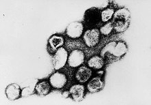
Rubella virus
| Rubella | |
|---|---|

| |
| Virus classification | |
| Group: | Group IV ((+)ssRNA)
|
| Family: | |
| Genus: | |
| Species: | Rubella virus
|
- This page is for the virus. For the disease, see Rubella.
Rubella virus is the pathogenic agent of the disease Rubella, and is the cause of congenital rubella syndrome when infection occurs during the first weeks of pregnancy. Humans are the only known host of this virus.
Rubella virus is the only member of the genus of Rubivirus and belongs to the family of Togaviridae, whose members commonly have a genome of single-stranded RNA of positive polarity which is enclosed by an icosahedral capsid. The RNA-genome inside the capsid has a length of approximately 9'757 nucleotides and encodes for two non-structural as well as three structural proteins.[1] The capsid protein and the two glycosylated envelope proteins E1 and E2 make up for the three structural proteins.
The molecular basis for the causation of congenital rubella syndrome are not yet completely clear, but in vitro studies with cell lines showed that Rubella virus has an apoptotic effect on certain cell types. There is evidence for a p53-dependent mechanism.[2]
Structure
The spherical virus particles (virions) of Togaviridae have a diameter of 50 to 70 nm and are covered by a lipid membrane (viral envelope), derived from the host cell membrane. There are prominent "spikes" (projections) of 6 nm composed of the viral envelope proteins E1 and E2 embedded in the membrane.[3]
Inside the lipid envelope is a capsid of 40 nm in diameter.
Replication
Togaviruses attach to the cell surface via specific receptors and are taken up by an endosome being formed. At the neutral pH outside of the cell the E2 envelope protein covers the E1 protein. The dropping pH inside the endosome frees the outer domain of E1 and causes the fusion of the viral envelope with the endosomal membrane. Thus, the capsid reaches the cytosol, decays and releases the genome
The (+)ssRNA (positive, singlestranded RNA) at first only acts as a template for the translation of the non-structural proteins, which are synthesized as a large polyprotein and are then cut into single proteins. The sequences for the structural proteins are first replicated by the viral RNA polymerase (Replicase) via a complementary (-)ssRNA as a template and translated as a separate short mRNA. This short subgenomic RNA is additionally packed in a virion. [4]
Translation of the structural proteins also produces a large polypeptide (110 Dalton). This is then endoproteolytically cut into E1, E2 and the capsid protein. E1 and E2 are type I transmembrane proteins which are transported into the endoplasmatic reticulum (ER) with the help of an N-terminal signal sequence. From the ER the heterodimeric E1·E2-complex reaches the Golgi apparatus, where the budding of new virions occurs (unlike alpha viruses, where budding occurs at the plasma membrane. The capsid proteins on the other hand stay in the cytoplasm and interact with the genomic RNA, together forming the capsid.[5]
Capsid protein
The capsid protein (protein C) has different functions. Its main tasks are the formation of homooligomeres to form the capsid, and the binding of the genomic RNA. Further is it responsible for the aggregation of RNA in the capsid, it interacts with the membrane proteins E1 and E2 and binds the human host-protein p32 which is important for replication of the virus in the host.[6]
As opposed to alpha viruses the capsid does not undergo autoprotolysis, rather is it cut off from the rest of the polyprotein by the signal-peptidase. Production of the capsid happens at the surface of intracellular membranes simultaneously with the budding of the virus.[7]
Literature
- David M. Knipe, Peter M. Howley et al. (eds.): Fields´ Virology 4. Auflage, Philadelphia 2001
- C.M. Fauquet, M.A. Mayo et al.: Eighth Report of the International Committee on Taxonomy of Viruses, London San Diego 2005
References
- ^ Dominguez G, Wang CY, Frey TK (1990). "Sequence of the genome RNA of rubella virus: evidence for genetic rearrangement during togavirus evolution". Virology. 177 (1): 225–38. PMID 2353453.
{{cite journal}}: Unknown parameter|month=ignored (help)CS1 maint: multiple names: authors list (link) - ^ Megyeri K, Berencsi K, Halazonetis TD; et al. (1999). "Involvement of a p53-dependent pathway in rubella virus-induced apoptosis". Virology. 259 (1): 74–84. doi:10.1006/viro.1999.9757. PMID 10364491.
{{cite journal}}: Explicit use of et al. in:|author=(help); Unknown parameter|month=ignored (help)CS1 maint: multiple names: authors list (link) - ^ Bardeletti G, Kessler N, Aymard-Henry M (1975). "Morphology, biochemical analysis and neuraminidase activity of rubella virus". Arch. Virol. 49 (2–3): 175–86. PMID 1212096.
{{cite journal}}: CS1 maint: multiple names: authors list (link) - ^ "Togaviridae- Classification and Taxonomy".
- ^ Garbutt M, Law LM, Chan H, Hobman TC (1999). "Role of rubella virus glycoprotein domains in assembly of virus-like particles". J. Virol. 73 (5): 3524–33. PMC 104124. PMID 10196241.
{{cite journal}}: Unknown parameter|month=ignored (help)CS1 maint: multiple names: authors list (link) - ^ Beatch MD, Everitt JC, Law LJ, Hobman TC (2005). "Interactions between rubella virus capsid and host protein p32 are important for virus replication". J. Virol. 79 (16): 10807–20. doi:10.1128/JVI.79.16.10807-10820.2005. PMC 1182682. PMID 16051872.
{{cite journal}}: Unknown parameter|month=ignored (help)CS1 maint: multiple names: authors list (link) - ^ Beatch MD, Hobman TC (2000). "Rubella virus capsid associates with host cell protein p32 and localizes to mitochondria". J. Virol. 74 (12): 5569–76. PMC 112044. PMID 10823864.
{{cite journal}}: Unknown parameter|month=ignored (help)
