Bacillary angiomatosis
| Bacillary angiomatosis | |
|---|---|
 | |
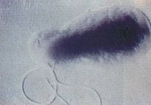
| Bartonella (bacterial species which causes this condition) | |
| Specialty | Infectious diseases |
Bacillary angiomatosis (BA) is a form of angiomatosis associated with bacteria of the genus Bartonella.[1]
Symptoms
Cutaneous BA is characterised by the presence of lesions on or under the skin. Appearing in numbers from one to hundreds, these lesions may take several forms:[citation needed]
- papules or nodules which are red, globular and non-blanching, with a vascular appearance
- purplish nodules sufficiently similar to Kaposi's sarcoma that a biopsy may be required to verify which of the two it is
- a purplish lichenoid plaque
- a subcutaneous nodule which may have ulceration, similar to a bacterial abscess
While cutaneous BA is the most common form, it can also affect several other parts of the body, such as the brain, bone, bone marrow, lymph nodes, gastrointestinal tract, respiratory tract, spleen, and liver. Symptoms vary depending on which parts of the body are affected; for example, those whose livers are affected may have an enlarged liver and fever, while those with osseous BA experience intense pain in the affected area.[citation needed]
Presentation
BA is characterised by the proliferation of blood vessels, resulting in them forming tumour-like masses in the skin and other organs.[citation needed]
Causes
It is caused by either Bartonella henselae or B. quintana.[2]
- B. henselae is most often transmitted through a cat scratch or bite,[3] though ticks and fleas may also act as vectors.
- B. quintana is usually transmitted by lice.
It can manifest in people with AIDS[4] and rarely appears in those who are immunocompetent.[citation needed]
Diagnosis
Diagnosis is based on a combination of clinical features and biopsy.
Neutrophilic infiltrate.[1]
Treatment and prevention
While curable, BA is potentially fatal if not treated.[1] BA responds dramatically to several antibiotics. Usually, erythromycin will cause the skin lesions to gradually fade away in the next four weeks, resulting in complete recovery. Doxycycline may also be used. However, if the infection does not respond to either of these, the medication is usually changed to tetracycline. If the infection is serious, then a bactericidal medication may be coupled with the antibiotics[citation needed]
If a cat is carrying Bartonella henselae, then it may not exhibit any symptoms. Cats may be bacteremic for weeks to years, but infection is more common in young cats. Transmission to humans is thought to occur via flea feces inoculated into a cat scratch or bite, and transmission between cats occurs only in the presence of fleas. Therefore, elimination and control of fleas in the cat's environment are key to prevention of infection in both cats and humans.[citation needed]
History
The condition that later became known as bacillary angiomatosis was first described by Stoler and associates in 1983.[5] Being unaware of its infectious origin, it was originally called epithelioid angiomatosis.[6] Following documentation of bacilli in Warthin-Starry stains and by electron microscopy in a series of cases by LeBoit and colleagues, the term bacillary angiomatosis was widely adopted.[7]
See also
References
- ^ a b c "Bacillary Angiomatosis: Practice Essentials, Background, Pathophysiology". 2017-02-09.
{{cite journal}}: Cite journal requires|journal=(help) - ^ Koehler JE, Sanchez MA, Garrido CS, et al. (December 1997). "Molecular epidemiology of bartonella infections in patients with bacillary angiomatosis-peliosis". N. Engl. J. Med. 337 (26): 1876–83. doi:10.1056/NEJM199712253372603. PMID 9407154.
- ^ Mateen FJ, Newstead JC, McClean KL (July 2005). "Bacillary angiomatosis in an HIV-positive man with multiple risk factors: A clinical and epidemiological puzzle". Can J Infect Dis Med Microbiol. 16 (4): 249–52. doi:10.1155/2005/230396. PMC 2095030. PMID 18159553.
- ^ Gasquet S, Maurin M, Brouqui P, Lepidi H, Raoult D (1998). "Bacillary angiomatosis in immunocompromised patients". AIDS. 12 (14): 1793–803. doi:10.1097/00002030-199814000-00011. PMID 9792380. S2CID 38201766.
- ^ Stoler MH, Bonfiglio TA, Steigbigel RT, Pereira M (Nov 1983). "An atypical subcutaneous infection associated with acquired immune deficiency syndrome". Am J Clin Pathol. 80 (5): 714–8. doi:10.1093/ajcp/80.5.714. PMID 6637883.
{{cite journal}}: CS1 maint: multiple names: authors list (link) - ^ Cockerell CJ, Whitlow MA, Webster GF, Friedman-Kien AE (Sep 1987). "Epithelioid angiomatosis: a distinct vascular disorder in patients with the acquired immunodeficiency syndrome or AIDS-related complex". Lancet. 2 (8560): 654–6. doi:10.1016/s0140-6736(87)92442-1. PMID 2887942. S2CID 1581891.
{{cite journal}}: CS1 maint: multiple names: authors list (link) - ^ LeBoit PE, Berger TG, Egbert BM, Beckstead JH, Yen TS, Stoler MH (1989). "Bacillary angiomatosis. The histopathology and differential diagnosis of a pseudoneoplastic infection in patients with human immunodeficiency virus disease". Am J Surg Pathol. 13 (11): 909–20. doi:10.1097/00000478-198911000-00001. PMID 2802010. S2CID 25225247.
