Vimentin
Template:PBB Vimentin is a member of the intermediate filament family of proteins. Intermediate filaments are an important structural feature of eukaryotic cells. They, along with microtubules and actin microfilaments, make up the cytoskeleton. Although most intermediate filaments are stable structures, in fibroblasts, vimentin exists as a dynamic structure. This filament is used as a marker for mesodermally derived tissues, and as such can be used as an immunohistochemical marker for sarcomas.
Structure
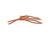
A vimentin monomer, like all other intermediate filaments, has a central α-helical domain, capped on each end by non-helical amino (head) and carboxy (tail) end domains.[1] Two monomers will twist around each other to form a coiled-coil dimer. Two dimers then form a tetramer, which, in turn, form a sheet by interacting with other tetramers.
The α-helical sequences contain a pattern of hydrophobic amino acids that contribute to forming a "hydrophobic seal" on the surface of the helix.[1] This seal allows the two helices to come together and coil. In addition, there is a periodic distribution of acidic and basic amino acids that seems to play an important role in stabilizing coiled-coil dimers.[1] The spacing of the charged residues is optimal for ionic salt bridges, which allows for the stabilization of the α-helix structure. While this type of stabilization is intuitive for intrachain interactions, rather than interchain interactions, scientists have proposed that perhaps the switch from intrachain salt bridges formed by acidic and basic residues to the interchain ionic associations contributes to the assembly of the filament.[1]
Function
Vimentin plays a significant role in supporting and anchoring the position of the organelles in the cytosol. Vimentin is attached to the nucleus, endoplasmic reticulum, and mitochondria, either laterally or terminally.[2] Vimentin Clips offers three different clips that show vimentin movement inside the cell.
The dynamic nature of vimentin is important when offering flexibility to the cell. Scientists found that vimentin provided cells with a resilience absent from the microtubule or actin filament networks, when under mechanical stress in vivo. Therefore, in general, it is accepted that vimentin is the cytoskeletal component responsible for maintaining cell integrity. (It was found that cells without vimentin are extremely delicate when disturbed with a micropuncture.) [3]
Results of a study involving transgenic mice that lacked vimentin[3] showed that the mice were functionally normal. While the outcome might seem surprising, it is possible that the microtubule network may have compensated for the absence of the intermediate network. This strengthens the suggestion of intimate interactions between microtubules and vimentin. Moreover, when microtubule depolymerizers were present, vimentin reorganization occurred, once again implying a relationship between the two systems.[3]
Vimentin Images offers a gallery of images in which vimentin and other cytoskeletal structures are labeled. These images allow the visualization of interactions between vimentin and other cytoskeletal components.
In essence, vimentin is responsible for maintaining cell shape, integrity of the cytoplasm, and stabilizing cytoskeletal interactions.
Also, vimentin is found to control the transport of low-density lipoprotein, LDL, -derived cholesterol from a lysosome to the site of esterification.[4] With the blocking of transport of LDL-derived cholesterol inside the cell, cells were found to store a much lower percentage of the lipoprotein than normal cells with vimentin. This dependence seems to be the first process of a biochemical function in any cell that depends on a cellular intermediate filament network. This type of dependence has ramifications on the adrenal cells, which rely on cholesteryl esters derived from LDL.[4]
Clinical significance
It has been used as a sarcoma tumor marker to identify mesenchyme.[5][6]
See also Anti-citrullinated protein antibody for its use in diagnosis of rheumatoid arthritis.
Interactions
Vimentin has been shown to interact with UPP1,[7] MYST2,[8][9] Desmoplakin,[10] Plectin,[11][12] SPTAN1,[12] MEN1,[13] Protein kinase N1[14] and YWHAZ.[15]
References
- ^ a b c d Fuchs E., Weber K. (1994). "Intermediate filaments: structure, dynamics, function, and disease". Annu Rev Biochem. 63: pp. 345–82. doi:10.1146/annurev.bi.63.070194.002021. PMID 7979242.
{{cite journal}}:|pages=has extra text (help) - ^ Katsumoto T., Mitsushima A., Kurimura T. (1990). "The role of the vimentin intermediate filaments in rat 3Y1 cells elucidated by immunoelectron microscopy and computer-graphic reconstruction". Biol Cell. 68 (2): pp. 139–46. doi:10.1016/0248-4900(90)90299-I. PMID 2192768.
{{cite journal}}:|pages=has extra text (help)CS1 maint: multiple names: authors list (link) - ^ a b c Goldman R. D., Khuon S., Chou Y., Opal P., Steinert P. (1996). "The function of intermediate filaments in cell shape and cytoskeletal integrity". J Cell Biol. 134 (4): pp. 971–83. doi:10.1083/jcb.134.4.971. PMID 8769421.
{{cite journal}}:|pages=has extra text (help)CS1 maint: multiple names: authors list (link) - ^ a b Sarria A. J., Panini S. R., Evans R. M. (1992). "A functional role for vimentin intermediate filaments in the metabolism of lipoprotein-derived cholesterol in human SW-13 cells". J Biol Chem. 267 (27): pp. 19455–63. PMID 1527066.
{{cite journal}}:|pages=has extra text (help)CS1 maint: multiple names: authors list (link) - ^ Leader M, Collins M, Patel J, Henry K (1987). "Vimentin: an evaluation of its role as a tumour marker". Histopathology. 11 (1): 63–72. PMID 2435649.
{{cite journal}}: Unknown parameter|month=ignored (help)CS1 maint: multiple names: authors list (link) - ^ "Immunohistochemistry from the Washington Animal Disease Diagnostic laboratory (WADDL)of the College of Veterinary Medicine, Washington State University". Retrieved 2009-03-14.
- ^ Russell, R L (2001). "Uridine phosphorylase association with vimentin. Intracellular distribution and localization". J. Biol. Chem. 276 (16). United States: 13302–7. doi:10.1074/jbc.M008512200. ISSN 0021-9258. PMID 11278417.
{{cite journal}}: Check date values in:|year=(help); Cite has empty unknown parameters:|laydate=,|laysource=, and|laysummary=(help); Unknown parameter|coauthors=ignored (|author=suggested) (help); Unknown parameter|month=ignored (help); Unknown parameter|quotes=ignored (help)CS1 maint: unflagged free DOI (link) CS1 maint: year (link) - ^ Rual, Jean-François (2005). "Towards a proteome-scale map of the human protein-protein interaction network". Nature. 437 (7062). England: 1173–8. doi:10.1038/nature04209. PMID 16189514.
{{cite journal}}: Check date values in:|year=(help); Cite has empty unknown parameters:|laydate=,|laysource=, and|laysummary=(help); Unknown parameter|coauthors=ignored (|author=suggested) (help); Unknown parameter|month=ignored (help); Unknown parameter|quotes=ignored (help)CS1 maint: year (link) - ^ Stelzl, Ulrich (2005). "A human protein-protein interaction network: a resource for annotating the proteome". Cell. 122 (6). United States: 957–68. doi:10.1016/j.cell.2005.08.029. ISSN 0092-8674. PMID 16179252.
{{cite journal}}: Check date values in:|year=(help); Cite has empty unknown parameters:|laydate=,|laysource=, and|laysummary=(help); Unknown parameter|coauthors=ignored (|author=suggested) (help); Unknown parameter|month=ignored (help); Unknown parameter|quotes=ignored (help)CS1 maint: year (link) - ^ Meng, J J (1997). "Two-hybrid analysis reveals fundamental differences in direct interactions between desmoplakin and cell type-specific intermediate filaments". J. Biol. Chem. 272 (34). UNITED STATES: 21495–503. ISSN 0021-9258. PMID 9261168.
{{cite journal}}: Check date values in:|year=(help); Cite has empty unknown parameters:|laydate=,|laysource=, and|laysummary=(help); Unknown parameter|coauthors=ignored (|author=suggested) (help); Unknown parameter|month=ignored (help); Unknown parameter|quotes=ignored (help)CS1 maint: year (link) - ^ Herrmann, H (1987). "Plectin and IFAP-300K are homologous proteins binding to microtubule-associated proteins 1 and 2 and to the 240-kilodalton subunit of spectrin". J. Biol. Chem. 262 (3). UNITED STATES: 1320–5. ISSN 0021-9258. PMID 3027087.
{{cite journal}}: Check date values in:|year=(help); Cite has empty unknown parameters:|laydate=,|laysource=, and|laysummary=(help); Unknown parameter|coauthors=ignored (|author=suggested) (help); Unknown parameter|month=ignored (help); Unknown parameter|quotes=ignored (help)CS1 maint: year (link) - ^ a b Brown, M J (2001). "Cutting edge: integration of human T lymphocyte cytoskeleton by the cytolinker plectin". J. Immunol. 167 (2). United States: 641–5. ISSN 0022-1767. PMID 11441066.
{{cite journal}}: Check date values in:|year=(help); Cite has empty unknown parameters:|laydate=,|laysource=, and|laysummary=(help); Unknown parameter|coauthors=ignored (|author=suggested) (help); Unknown parameter|month=ignored (help); Unknown parameter|quotes=ignored (help)CS1 maint: year (link) - ^ Lopez-Egido, Juan (2002). "Menin's interaction with glial fibrillary acidic protein and vimentin suggests a role for the intermediate filament network in regulating menin activity". Exp. Cell Res. 278 (2). United States: 175–83. ISSN 0014-4827. PMID 12169273.
{{cite journal}}: Check date values in:|year=(help); Cite has empty unknown parameters:|laydate=,|laysource=, and|laysummary=(help); Unknown parameter|coauthors=ignored (|author=suggested) (help); Unknown parameter|month=ignored (help); Unknown parameter|quotes=ignored (help)CS1 maint: year (link) - ^ Matsuzawa, K (1997). "Domain-specific phosphorylation of vimentin and glial fibrillary acidic protein by PKN". Biochem. Biophys. Res. Commun. 234 (3). UNITED STATES: 621–5. doi:10.1006/bbrc.1997.6669. ISSN 0006-291X. PMID 9175763.
{{cite journal}}: Check date values in:|year=(help); Cite has empty unknown parameters:|laydate=,|laysource=, and|laysummary=(help); Unknown parameter|coauthors=ignored (|author=suggested) (help); Unknown parameter|month=ignored (help); Unknown parameter|quotes=ignored (help)CS1 maint: year (link) - ^ Tzivion, G (2000). "Calyculin A-induced vimentin phosphorylation sequesters 14-3-3 and displaces other 14-3-3 partners in vivo". J. Biol. Chem. 275 (38). UNITED STATES: 29772–8. doi:10.1074/jbc.M001207200. ISSN 0021-9258. PMID 10887173.
{{cite journal}}: Check date values in:|year=(help); Cite has empty unknown parameters:|laydate=,|laysource=, and|laysummary=(help); Unknown parameter|coauthors=ignored (|author=suggested) (help); Unknown parameter|month=ignored (help); Unknown parameter|quotes=ignored (help)CS1 maint: unflagged free DOI (link) CS1 maint: year (link)


